Acta medica Lituanica ISSN 1392-0138 eISSN 2029-4174
2026. Vol. 33. No 2. Online ahead of print DOI: https://doi.org/10.15388/Amed.2026.33.2.9
Orhan Ay*
Perinatology Department, Medical Faculty, Necmettin Erbakan University, Konya, Turkey
E-mail: orhan.ay.5561@gmail.com
ORCID ID https://orcid.org/0000-0002-9685-5374
Fethiye Şahin
Perinatology Department, Medical Faculty, Necmettin Erbakan University, Konya, Turkey
E-mail: drfethiye@gmail.com
ORCID ID https://orcid.org/0009-0007-9817-9481
Pelin Bahçeci
Department of Obstetrics and Gynecology, Faculty of Medicine, Necmettin Erbakan University, Konya, Turkey
E-mail: pelinbahceci30@gmail.com
ORCID ID https://orcid.org/0009-0008-2615-3052
Hasan Madenci
Department of Pediatric Surgery, Faculty of Medicine, Necmettin Erbakan University, Konya, Turkey
E-mail: hasanmadenci@gmail.com
ORCID ID https://orcid.org/0000-0002-7243-5340
Ali Acar
Perinatology Department, Medical Faculty, Necmettin Erbakan University, Konya, Turkey
E-mail: mdaliacar@gmail.com
ORCID ID https://orcid.org/0009-0006-1474-3958
Abstract. Background: Sacrococcygeal teratoma (SCT) is the most common congenital neoplasm in fetuses and newborns. Altman Type III lesions may be difficult to diagnose and manage prenatally because of their marked intrapelvic extension.
Materials and Methods: A 24-year-old primigravida woman was referred at 20 weeks of gestation after routine ultrasonography revealed a sacrococcygeal mass extending into the abdomen. Prenatal evaluation included ultrasonography, fetal magnetic resonance imaging, and chromosomal microarray analysis. The pregnancy was managed by a multidisciplinary team with serial fetal surveillance.
Results: Imaging demonstrated a predominantly cystic lesion which was consistent with Altman Type III SCT. During the follow-up, bilateral fetal hydronephrosis, gestational diabetes mellitus, and mild polyhydramnios developed. At 38 weeks, an elective cesarean section was performed. On postnatal day 4, total tumor resection with coccygectomy was successfully completed. Histopathology confirmed a mature teratoma. At 6 months, persistent hydronephrosis remained under follow-up, with no documented recurrence.
Conclusions: Accurate prenatal imaging, close surveillance, multidisciplinary planning, and timely postnatal surgery are essential for favorable outcomes in Type III fetal SCT.
Keywords: sacrococcygeal teratoma, fetal MRI, prenatal diagnosis, hydronephrosis, coccygectomy.
Santrauka. Įvadas: Sakrokoksigealinė teratoma (SCT) yra dažniausias vaisiaus ir naujagimio įgimtas navikas. Altmano III tipo pažeidimus gali būti sunku diagnozuoti ir gydyti prieš gimimą dėl jų ryškaus išplitimo į dubens ertmę.
Medžiaga ir metodai: 24 metų pirmą kartą nėščiai moteriai 20-ąją nėštumo savaitę, atliekant įprastą ultragarsinį tyrimą, buvo nustatytas į pilvo ertmę išsiplėtęs sakrokoksigealinis navikas. Prenatalinis vertinimas apėmė ultragarsinį tyrimą, vaisiaus magnetinio rezonanso tomografiją ir chromosomų kopijų skaičiaus tyrimą. Nėštumą prižiūrėjo daugiadisciplinė komanda, atliekanti nuolatinę vaisiaus stebėseną.
Rezultatai: Vaizdo tyrimai parodė daugiausia cistinį pažeidimą, atitinkantį Altmano III tipo SCT. Stebėjimo metu išsivystė abipusė vaisiaus hidronefrozė, gestacinis diabetas ir lengvas polihidramnionas. Moteriai 38-ąją nėštumo savaitę buvo atliktas planinis cezario pjūvis. Ketvirtą dieną po gimdymo sėkmingai atlikta visiška naviko rezekcija kartu pašalinant uodegikaulį. Histopatologinis tyrimas patvirtino subrendusią teratomą. Po 6 mėnesių buvo matoma išlikusi hidronefrozė, tačiau atsinaujinimo atvejų nebuvo užfiksuota.
Išvados: Tikslus prenatalinis vaizdinis tyrimas, atidus stebėjimas, daugiadisciplinis planavimas ir laiku atlikta operacija po gimdymo yra būtini siekiant palankių rezultatų III tipo vaisiaus SCT atveju.
Raktiniai žodžiai: sakrokoksigealinė teratoma, vaisiaus MRT, prenatalinė diagnostika, hidronefrozė, kokcigektomija.
________
* Corresponding author
Received: 24/10/2025. Revised: 13/04/2026. Accepted: 23/06/2026
Copyright © 2026 Orhan Ay, Fethiye Şahin, Pelin Bahçeci, Hasan Madenci, Ali Acar. Published by Vilnius University Press.This is an Open Access article distributed under the terms of the Creative Commons Attribution License, which permits unrestricted use, distribution, and reproduction in any medium, provided the original author and source are credited.
Sacrococcygeal Teratoma (SCT) is a congenital tumor which develops due to the abnormal proliferation of totipotent germ cells at Hensen’s node during the embryonic period. It is the most common congenital neoplasm in fetuses and newborns, with an estimated incidence of approximately 1 in 27,000 live births, and more than 75% of cases occur in female fetuses [1,2]. Histologically, sacrococcygeal teratomas are classified as mature, immature, and malignant forms. The mature type contains well-differentiated tissues, whereas the immature type is characterized by the predominance of neural elements and tends to exhibit a more aggressive clinical course [3].
According to the classification proposed by the Surgical Section of the American Academy of Pediatrics, sacrococcygeal teratomas are divided into four types: Type I is entirely external, Type II is predominantly external with an intrapelvic extension, Type III has both external and significant intrapelvic components that are closely related to pelvic organs, and Type IV is completely intrapelvic in location [4].
Ultrasonography (USG) is the primary diagnostic modality in the prenatal detection of sacrococcygeal teratomas, as it enables visualization of both cystic and solid components, allowing early identification of the lesion [5]. However, particularly in Type III and Type IV cases, there are limitations in assessing the intrapelvic extension of the mass and its relationship with the surrounding organs. Therefore, fetal Magnetic Resonance Imaging (MRI) provides valuable information for delivery planning by clearly demonstrating the external and internal components of the lesion through its superior soft-tissue contrast resolution.
Prenatal diagnosis is most commonly established during routine ultrasonographic evaluation in the second trimester, although first-trimester diagnoses have also been reported [6,7]. Most diagnosed cases of sacrococcygeal teratoma (SCT) are classified as Altman Type I or Type II [8]. In the experience of the United Kingdom Children’s Cancer Study Group (UKCCSG) involving 37 cases of sacrococcygeal teratoma (SCT), the distribution of Altman Type I, II, III, and IV lesions was 62%, 14%, 19%, and 6%, respectively [9]. Type I, II, and III tumors grow as exophytic masses and can therefore be externally visible. These tumor types are more easily diagnosed both in the prenatal and neonatal periods and are generally associated with a lower malignant potential [10]. On ultrasonography, sacrococcygeal teratomas typically appear as a mass located near the distal spine. Due to their mass effect, they may cause bladder outlet obstruction, hydronephrosis, rectal stenosis or atresia, vascular shunting, and cardiomegaly secondary to high-output cardiac failure [11].
The prognosis of prenatally diagnosed cases is closely related to the tumor’s size, vascularity, proportion of solid components, and associated cardiac load. In large and highly vascular masses, the development of high-output cardiac failure and hydrops fetalis significantly increases fetal and neonatal mortality [12].
When deciding on the mode of delivery, tumor size, morphological characteristics, vascularity, and the accompanying obstetric conditions should all be taken into consideration. Although vaginal delivery may be feasible in small, low-risk lesions, particularly those that are predominantly cystic, elective cesarean section is more commonly preferred in large, protruding, or markedly solid/vascular masses in order to reduce the risks of dystocia, tumor rupture, and hemorrhage [13]. Prenatal multidisciplinary planning facilitates delivery-room preparedness and coordinated postnatal management, and careful prenatal monitoring with individualized treatment has been associated with improved neonatal outcomes [13].
In the postnatal period, recurrence remains an important clinical concern; it has been reported in approximately 10%–12% of cases in recent series [14,15]. The risk appears to be higher in tumors with immature or malignant histology, and earlier studies have additionally linked recurrence to incomplete resection and omission of coccygectomy [16]. Therefore, complete excision with coccygectomy remains a key component of standard surgical management.
Advances in prenatal ultrasonography and fetal MRI have substantially improved the antenatal detection and characterization of sacrococcygeal teratomas, thereby supporting more accurate counseling, delivery planning, and postnatal management [17]. These advancements allow potential complications to be anticipated during the prenatal period and enable the individualization of postnatal treatment strategies. In this context, the present case report discusses the prenatal diagnostic process, delivery management, and postnatal clinical course of a fetus diagnosed with an Altman Type III sacrococcygeal teratoma during the prenatal period, in light of current literature.
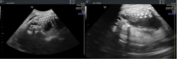
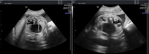
A 24-year-old primigravida woman was referred to our clinic at 20 weeks of gestation after a routine detailed ultrasound examination revealed a mass located in the sacrococcygeal region extending into the abdomen. Ultrasonography demonstrated a 30 × 35 mm predominantly cystic lesion originating from the sacrococcygeal area and extending toward the bladder, containing minimal solid components. No significant vascularity was observed within the mass. Hydrops, cardiomegaly, or pericardial effusion were not detected. Apart from this finding, no other structural fetal anomalies were identified. Amniocentesis was performed, and chromosomal microarray analysis was requested. The genetic evaluation was reported as normal. Because of the lesion’s intrapelvic extension, an intra-abdominal cystic mass was also considered in the differential diagnosis (Figure 1). Compression of the bladder was evident on ultrasonography (Figure 2).
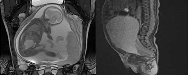
This finding made the diagnosis somewhat challenging. The mass located in the sacrococcygeal region and its connection with the intrapelvic area were evaluated as being consistent with an Altman Type III sacrococcygeal teratoma. Fetal Magnetic Resonance Imaging (MRI) was performed with the objective to further clarify the nature and extent of the lesion (Figure 3). The mass was located posteriorly in the gluteal region and extended anteriorly to the posterior aspect of the bladder.
The pregnancy was managed by a multidisciplinary team including specialists in perinatology, neonatology, anesthesiology, and pediatric surgery. Fetal growth was assessed every 2–3 weeks, while amniotic fluid volume, umbilical artery Doppler, and middle cerebral artery Doppler measurements were performed regularly, each week. From 32 weeks onward, nonstress testing was initiated on a weekly basis.
During the follow-up, bilateral fetal hydronephrosis developed secondary to bladder compression caused by the teratoma. At 26 weeks of gestation, a 75 g oral glucose tolerance test was performed, and the patient was diagnosed with gestational diabetes mellitus. Blood glucose levels remained well-controlled with dietary management, and the pregnancy continued uneventfully. After 26 weeks, mild polyhydramnios developed, with an amniotic fluid index of 28 cm. No additional fetal abnormalities were detected during the follow-up. At 38 weeks of gestation, an elective cesarean section was performed, delivering a live male infant weighing 3210 g with Apgar scores of 8 and 9 (Figure 4).
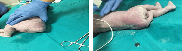
At the initial postnatal examination, a lobulated, elastic mass was observed in the sacrococcygeal region. Postnatal MRI demonstrated a 53 × 90 mm cystic lesion with high internal signal intensity occupying the abdominal cavity, displacing both kidneys laterally and bowel loops anterolaterally. No involvement of the bladder wall was detected. The abdominal portion of the lesion was predominantly cystic, whereas solid components were located in the pelvic and sacrococcygeal regions. On the fourth postnatal day, total tumor resection and coccygectomy were successfully performed (Figure 5).
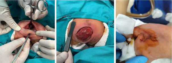
Regarding the surgical technique, the neonate was initially placed in the prone position, and the sacrococcygeal component was exposed through an inverted V (chevron) incision. Sharp and blunt dissection was performed with careful preservation of the anal canal and rectal integrity. The pelvic component was found to be attached to the coccyx, and coccygectomy was performed during the resection. The sacrococcygeal portion of the mass was excised first. The patient was then repositioned supine, and a Pfannenstiel incision was made to allow excision of the intra-abdominal extension. The abdominal component was dissected free from the surrounding tissues and sigmoid mesentery, and complete tumor excision was achieved. Hemostasis was secured, and a Penrose drain was placed. Intraoperative blood loss was minimal, and no transfusion was required. The postoperative course was uneventful. Renal ultrasonography performed on postoperative day 5 revealed grade 3 hydronephrosis in both kidneys. The infant was discharged on postoperative day 10.
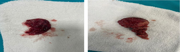
Histopathological examination confirmed a mature teratoma (Figure 6), demonstrating well-differentiated tissues derived from all three germ layers, with no immature or malignant components.
At 6 months of age, the infant was still being followed by the pediatric hematology and pediatric nephrology teams. Nephrology follow-up was maintained because of persistent hydronephrosis. Follow-up renal ultrasonography showed bilateral grade 3 hydronephrosis, with renal pelvic anteroposterior diameters of 12 mm on the right and 13 mm on the left, as well as mild bilateral cortical thinning; no definite bladder abnormality was identified within the limits of the examination. The infant had one documented urinary tract infection during the follow-up. Apart from this, the clinical course was unremarkable, and no recurrence was documented in the available records.
This case report has several limitations. First, it includes only a single patient; therefore, the findings cannot be generalized to all cases of sacrococcygeal teratoma. Second, the classification of the lesion as Altman Type III poses an additional diagnostic challenge, as the prominent intrapelvic extension and the relatively less conspicuous external component in this subtype may make prenatal differential diagnosis difficult when based on ultrasonography alone. Although fetal MRI helped to delineate the origin and extent of the lesion more clearly in our case, this diagnostic difficulty during prenatal assessment should still be considered an important limitation. Finally, although the follow-up data up to 6 months were available, longer-term outcomes could not yet be fully assessed. In particular, the persistence of hydronephrosis indicates the need for continued surveillance, and definitive evaluation of long-term renal and urinary function, bowel function, and recurrence risk requires a longer follow-up.
With the advancement of modern ultrasonography and fetal MRI techniques, the rate of prenatal diagnosis has markedly increased. However, Altman Type III and Type IV cases present a more complex clinical picture in terms of diagnosis and management, owing to their prominent pelvic extension. In our case, a lesion detected at 20 weeks of gestation, characterized by prominent intrapelvic extension and cystic components, was classified as an Altman Type III sacrococcygeal teratoma (SCT). According to the literature, Type III cases pose challenges in both prenatal imaging and surgical planning, as the external component is limited and the internal extension predominates [4]. In such cases, fetal magnetic resonance imaging (MRI) assists diagnosis by providing high-resolution soft-tissue contrast, allowing clearer assessment of the tumor’s relationship with the bladder.
Studies have shown that cases with a high tumor-volume-to-fetal-weight ratio (TFR) along with marked hypervascularity exhibit a significantly increased mortality rate due to cardiac overload. Similarly, the presence of fetal hydrops and cardiomegaly has been identified as major risk factors associated with poor prognosis [18]. In our case, the absence of signs of hydrops or cardiac failure allowed the pregnancy to remain stable and to be carried successfully to 38 weeks of gestation.
In delivery management, recent reviews emphasize that no universally accepted protocol exists, and that the timing and mode of delivery should be tailored according to the tumor characteristics, fetal condition, and the experience and resources of the multidisciplinary team [13,19]. Elective cesarean delivery is often preferred in reported management algorithms, although vaginal delivery may still be feasible in selected small, low-risk tumors [19]. In our case, an elective cesarean section was performed, followed by early postnatal total tumor resection with coccygectomy. In the postnatal period, recurrence remains an important concern, and it has been reported in approximately 10%–12% of cases in recent multicenter series [14,15]. The risk appears to be higher in tumors with immature or malignant histology and after incomplete resection, while earlier studies have also implicated omission of coccygectomy [16]. Therefore, complete excision with coccygectomy remains a key component of standard surgical management [16].
Histopathological examination in our case confirmed the diagnosis of a mature teratoma. This finding is consistent with the majority of prenatally diagnosed sacrococcygeal teratoma cases reported in the literature, as most of these tumors are histologically benign, while malignant types are rare, accounting for approximately 12%–14% of the cases [17]. Nevertheless, immature sacrococcygeal teratomas have been reported to be associated with more rapid tumor growth, an increased risk of perinatal complications, a higher likelihood of recurrence, and greater mortality compared with mature forms [20].
Recent publications have begun to discuss fetal therapeutic approaches in the management of prenatally diagnosed sacrococcygeal teratomas. In severe cases complicated by hydrops, polyhydramnios, or high-output cardiac failure, intrauterine interventions such as drainage, vascular ablation, or the EXIT (Ex-Utero Intrapartum Treatment) procedure may be considered [13,19]. However, the outcomes of these interventions vary considerably according to the technique used and case selection, and survival rates of approximately 57%–61% have been reported in selected series [13,21].
Early and accurate imaging, comprehensive cardiac evaluation, multidisciplinary delivery planning, and complete postnatal surgical excision are essential for reducing mortality and morbidity in prenatally diagnosed Type III sacrococcygeal teratomas. Consistent with the current literature, this case further highlights the critical importance of meticulous fetal surveillance and the appropriate surgical timing in achieving favorable outcomes.
The authors declare that they have no conflict of interest regarding this study.
O. A.: conceptualization, formal analysis, writing – original draft preparation, writing – review and editing.
F. Ş.: data curation, methodology, visualization.
P. B.: data curation, validation.
H. M.: writing – review and editing.
A. A.: writing – review and editing.
Written informed consent for publication of this case and the accompanying images was obtained from the patient’s parents. As this report describes a single case presentation, ethical committee approval was not required.