Acta medica Lituanica ISSN 1392-0138 eISSN 2029-4174
2026. Vol. 33. No 2. Online ahead of print DOI: https://doi.org/10.15388/Amed.2026.33.2.7
Nitipong Siriwong
School of Science, Mae Fah Luang University, Thailand
E-mail: nitipong@mfu.ac.th
Ekachai Chukeatirote*
School of Science, Mae Fah Luang University, Thailand
E-mail: ekachai@mfu.ac.th
ORCID ID https://orcid.org/0000-0002-9968-5841
Abstract. Background: Staphylococcus aureus is a major opportunistic pathogen in hospitals, where sustained exposure to antimicrobial agents and disinfectants promotes the emergence and persistence of resistant strains. In addition, tolerance to heavy metals may facilitate co-selection and long-term survival of resistant S. aureus in clinical settings.
Purpose: This study investigated the occurrence of methicillin-resistant S. aureus (MRSA), vancomycin-intermediate S. aureus (VISA), and vancomycin-resistant S. aureus (VRSA), and evaluated antibiotic and heavy metal resistance profiles among S. aureus isolates recovered from hospitals.
Materials and Methods: Seventy-nine bacterial isolates were collected from three hospitals. Identification using Gram staining and biochemical tests identified 71 S. aureus isolates. Resistance to antibiotics was determined by disk diffusion, and was quantified by using the multiple antibiotic resistance (MAR) index. Resistance to heavy metals was assessed at increasing the concentrations, and the multiple heavy metal resistance (MHMR) index was then calculated. MRSA, VISA, and VRSA were screened by using selective agar containing oxacillin and vancomycin. Cluster analysis was performed based on combined antibiotic and heavy metal resistance profiles.
Results: High resistance rates were observed for penicillin and ampicillin (95.8% each), whereas most isolates showed low levels of resistance to chloramphenicol, bacitracin, streptomycin, kanamycin, and methicillin. MAR index values ranged from 0 to 0.88, with most isolates exceeding 0.2. All isolates exhibited resistance to multiple heavy metals, with MHMR values between 0.57 and 0.71, and marked tolerance to chromium and lead. Screening identified 9 MRSA, 3 VISA, and 1 VRSA isolate, including one strain classified as both MRSA and VRSA. Cluster analysis resolved nine major resistance-associated groups.
Conclusion: Hospital-associated S. aureus isolates showed extensive antibiotic and heavy metal resistance, supporting the likelihood of co-selection of resistance traits. These findings underscore the importance of integrated antimicrobial stewardship and environmental control strategies in hospitals.
Keywords: antibiotic resistance, heavy metal resistance, Staphylococcus.
Santrauka. Įvadas: Staphylococcus aureus yra vienas iš pagrindinių oportunistinių patogenų ligoninėse, kur nuolatinis sąlytis su antimikrobiniais preparatais ir dezinfekcinėmis priemonėmis skatina atsparių kamienų atsiradimą ir išlikimą. Be to, atsparumas sunkiesiems metalams gali palengvinti atsparių S. aureus kamienų bendrą atranką ir ilgalaikį išlikimą klinikinėje aplinkoje.
Tikslas: Šiame tyrime buvo tiriamas meticilinui atsparių S. aureus (MRSA), vidutinio atsparumo vankomicinui S. aureus (VISA) ir vankomicinui atsparių S. aureus (VRSA) paplitimas bei vertinami ligoninėse išskirtų S. aureus izoliatų atsparumo antibiotikams ir sunkiesiems metalams profiliai.
Medžiagos ir metodai: Iš trijų ligoninių buvo surinkta 79 bakterijų izoliatų. Atlikus identifikavimą naudojant Gramo dažymą ir biocheminius tyrimus, identifikuota 71 S. aureus izoliatas. Atsparumas antibiotikams buvo nustatytas disko difuzijos metodu, o jo laipsnis įvertintas naudojant daugialypio atsparumo antibiotikams (MAR) indeksą. Atsparumas sunkiesiems metalams buvo vertinamas didėjančiomis koncentracijomis, o daugialypio atsparumo sunkiesiems metalams (MHMR) indeksas buvo apskaičiuotas. MRSA, VISA ir VRSA atrinkti naudojant selektyvų agarą, kuriame buvo oksacilino ir vankomicino. Remiantis kombinuotais antibiotikų ir sunkiųjų metalų atsparumo profiliais buvo atlikta klasterių analizė.
Rezultatai: Nustatyti aukšti atsparumo penicilinui ir ampicilinui rodikliai (po 95,8 %), tuo tarpu daugumos izoliatų nustatytas žemas atsparumas chloramfenikoliui, bacitricinui, streptomicinui, kanamicinui ir meticilinui. MAR indekso vertės svyravo nuo 0 iki 0,88, daugumos izoliatų vertės viršijo 0,2. Visi izoliatai pasižymėjo atsparumu keliems sunkiesiems metalams, MHMR vertės buvo nuo 0,57 iki 0,71, taip pat buvo nustatytas didelis atsparumas chromui ir švinui. Atrankos metu identifikuoti 9 MRSA, 3 VISA ir 1 VRSA izoliatai, įskaitant vieną štamą, klasifikuotą kaip MRSA ir VRSA. Klasterių analizė leido išskirti devynias pagrindines atsparumo grupes.
Išvada: Su ligoninėmis susiję S. aureus izoliatai parodė didelį atsparumą antibiotikams ir sunkiųjų metalų poveikiui, o tai patvirtina atsparumo požymių bendros atrankos tikimybę. Šie rezultatai pabrėžia integruoto antimikrobinių vaistų valdymo ir aplinkos kontrolės strategijų svarbą ligoninėse.
Raktažodžiai: atsparumas antibiotikams, atsparumas sunkiesiems metalams, Staphylococcus.
__________
* Corresponding author
Received: 10/02/2026. Revised: 13/04/2026. Accepted: 01/07/2026
Copyright © 2026 Nitipong Siriwong, Ekachai Chukeatirote. Published by Vilnius University Press.This is an Open Access article distributed under the terms of the Creative Commons Attribution License, which permits unrestricted use, distribution, and reproduction in any medium, provided the original author and source are credited.
Staphylococcus aureus is a major opportunistic pathogen capable of causing a wide range of community- and hospital-acquired infections [1]. Such infections range from mild skin and wound infections to severe and life-threatening conditions such as septicemia, pneumonia, and endocarditis [2]. The clinical management of S. aureus infections has become increasingly difficult due to its exceptional ability to acquire resistance to multiple antimicrobial agents. Of particular concern is methicillin-resistant Staphylococcus aureus (MRSA), which has emerged as a significant global public health threat [3]. MRSA was first reported in England in 1961 [4], and, since then, its prevalence has increased worldwide, posing a major challenge to healthcare systems. Numerous MRSA outbreaks have been documented across different regions, including Southeast Asia [5]. MRSA strains can be broadly classified based on their epidemiology into hospital-associated MRSA (HA-MRSA), community-associated MRSA (CA-MRSA), and livestock-associated MRSA (LA-MRSA). These groups differ in clinical manifestations, molecular characteristics, transmission dynamics, and antibiotic susceptibility profiles [6]. The emergence of LA-MRSA has further raised concerns regarding zoonotic transmission and the potential spread of resistant strains to human populations.
Methicillin resistance in S. aureus is primarily mediated by the mecA gene, which is carried on a mobile genetic element known as the staphylococcal cassette chromosome mec (SCCmec) [7]. The mecA gene encodes a low-affinity penicillin-binding protein (PBP2a), conferring resistance to virtually all β-lactam antibiotics, including penicillins, cephalosporins, carbapenems, and flucloxacillin [8]. Consequently, glycopeptide antibiotics such as vancomycin and teicoplanin have been widely used as last-resort treatments for MRSA infections [9]. Alarmingly, the emergence of vancomycin-intermediate and vancomycin-resistant S. aureus (VISA and VRSA) has been reported, thus further complicating treatment options and raising serious public health concerns [10].
Beyond antibiotic exposure, the use of antiseptics, disinfectants, and heavy metals in healthcare and environmental settings has been increasingly recognized as an important factor contributing to the selection and persistence of resistant bacterial populations [11,12]. Heavy metals such as silver, copper, and zinc are commonly incorporated into medical devices, coatings, and antimicrobial formulations due to their biocidal properties, whereas metals such as chromium and lead are more frequently associated with industrial activities and environmental contamination [12]. The persistence of these metals in healthcare and surrounding environments may contribute to long-term selective pressure on microbial populations. Numerous studies have reported that resistance determinants for antibiotics and heavy metals are often co-located on mobile genetic elements, facilitating co-selection and maintenance of multidrug resistance even in the absence of antibiotic pressure [13,14].
In Thailand, MRSA has been recognized for more than four decades [15], with several studies documenting outbreaks in hospital settings [16-18]. Recent national surveillance reports have highlighted the continued burden of MRSA in the country [5,19]. However, data on the combined occurrence of antibiotic resistance and heavy metal tolerance among S. aureus isolates remain limited, particularly at the regional level. Therefore, this study was undertaken as part of an antibiotic resistance surveillance program in Chiang Rai, Thailand. The objectives were to (i) identify and characterize S. aureus clinical isolates from hospital settings; (ii) determine the prevalence of MRSA by using oxacillin-based screening; (iii) assess antibiotic resistance patterns and multiple antibiotic resistance (MAR) indices; (iv) evaluate heavy metal resistance profiles and multiple heavy metal resistance (MHMR) indices; and (v) analyze phenotypic relationships among isolates using cluster analysis based on combined antibiotic and heavy metal resistance profiles. The findings provide insight into resistance dynamics in hospital-associated S. aureus and highlight the potential role of environmental selective pressures in shaping antimicrobial resistance.
A total of 80 bacterial isolates were included in this study: 63 from Hospital A, 10 from Hospital B, and 6 from Hospital C. One laboratory strain, Staphylococcus aureus TISTR 1466, was included as a non-clinical susceptible control strain. The isolates were originally recovered from clinical specimens collected during routine diagnostic procedures in hospital microbiology laboratories between 2021 and 2022. For this study, isolates were obtained as anonymized stock cultures from hospital staff. No patient-identifiable information or clinical data were available to the researchers; therefore, the study did not involve human subjects. All isolates were stored on nutrient agar slants at 4 °C for short-term use and preserved at -70 °C in nutrient broth containing 20% glycerol. The isolates were coded according to their source as CRP (Hospital A), CRO (Hospital B), CRK (Hospital C), and TISTR 1466.
All isolates were initially cultured on nutrient agar and incubated at 37 °C for 24 h. Preliminary identification was performed by Gram staining and catalase testing. Gram-positive cocci arranged in clusters and exhibiting positive catalase reactions were considered presumptive Staphylococcus species. Further species-level identification was carried out by using standard biochemical tests, including hemolysis on blood agar (Oxoid Ltd., Basingstoke, UK), a coagulase test (Sigma-Aldrich, St. Louis, USA), and carbohydrate fermentation assays [20]. Glucose and mannitol fermentation were assessed by using phenol red broth, with changes in color indicating positive fermentation. Isolates showing β-hemolysis, positive coagulase reaction, and the ability to ferment glucose and mannitol were identified as Staphylococcus aureus. Isolates not fulfilling all identification criteria were excluded from subsequent analyses.
Antimicrobial susceptibility testing was performed using the disk diffusion method according to CLSI M100-S29 (2019) guidelines [21]. In brief, bacterial suspensions were adjusted to 0.5 McFarland standard and inoculated onto Mueller–Hinton agar (MHA: Oxoid, Basingstoke, UK). Antibiotic discs included ampicillin (10 µg), bacitracin (10 U), chloramphenicol (30 µg), erythromycin (15 µg), kanamycin (30 µg), methicillin (5 µg), penicillin G (10 U), streptomycin (10 µg), and tetracycline (30 µg) (all from Oxoid, Basingstoke, UK). Plates were incubated at 37 °C for 24 h, and inhibition zones were measured. Isolates were classified as resistant, intermediate, or susceptible based on CLSI criteria, wherever available. For antibiotics without CLSI breakpoints for S. aureus (e.g., ampicillin, kanamycin, streptomycin, bacitracin) and methicillin (no longer recommended for routine testing and replaced by cefoxitin as a surrogate marker), interpretation was based on previously published surveillance studies and consistent inhibition zone distribution patterns within the dataset [22]. The multiple antibiotic resistance (MAR) index was calculated for each S. aureus isolate by using the following formula: MAR index = a/b where a represents the number of antibiotics to which the isolate was resistant, and b represents the total number of antibiotics tested. MAR index values greater than 0.2 were considered indicative of exposure to environments with frequent antibiotic use and possible selective pressure [23].
Screening for phenotypically methicillin-resistant Staphylococcus aureus (MRSA) was performed by using oxacillin, following the method described by Brown et al. [24]. Briefly, bacterial suspensions were prepared and adjusted to approximately 10⁴ CFU/mL. Aliquots (10 µL) of each suspension were spot-inoculated onto MHA plates supplemented with 4% NaCl and 6 µg ml-1 oxacillin. The plates were incubated at 30 °C for 24 h, and visible bacterial growth was interpreted as indicative of MRSA. Screening for phenotypically vancomycin-intermediate S. aureus (VISA) and phenotypically vancomycin-resistant S. aureus (VRSA) was conducted by using vancomycin screen agar, as described by Gitanjali et al. [25]. Briefly, 10 µL aliquots of bacterial suspensions (approximately 10⁴ CFU mL-1) were spot-inoculated onto MHA plates containing vancomycin. Isolates exhibiting growth on plates containing 6 µg mL-1 vancomycin were classified as VISA, whereas growth on plates containing 16 µg mL-1 vancomycin was interpreted as VRSA.
Heavy metal resistance was assessed using MHA supplemented with seven heavy metals: AgNO3, CoCl2·6H2O, CuSO4·5H2O, HgCl2, K2Cr2O7, Pb(CH3COO)2·3H2O, and ZnSO4·7H2O (Merck, Darmstadt, Germany). Stock solutions of each compound were prepared and added to the medium with the objective to obtain final concentrations expressed in millimolar (mM). The concentration ranges used were 0.1, 0.5, 1, 5, 20, 40, and 80 mM, depending on the compound. Isolates were considered resistant when growth occurred at or above the defined threshold concentrations, namely, 1 mM for AgNO₃, CoCl₂·6H₂O, CuSO₄·5H₂O, K₂Cr₂O₇, Pb(CH₃COO)₂·3H₂O, and ZnSO₄·7H₂O, and 0.1 mM for HgCl₂, as previously described [26]. The reported concentrations refer to the nominal final concentrations of the added heavy metal salts in the culture medium. It is acknowledged that the actual bioavailability of metal ions may vary due to interactions with components of the growth medium, including complexation, precipitation, and changes in ionic equilibrium. The multiple heavy metal resistance (MHMR) index was calculated by using the following formula: MHMR index = a/b, where a represents the number of heavy metals to which the isolate was resistant, and b represents the total number of heavy metals tested. This index was used as a study-specific, descriptive metric and is not a standard index.
Phenotypic data obtained from antibiotic and heavy metal resistance assays were scored in a binary format, where resistance was assigned a value of ‘1’ and susceptibility was assigned a value of ‘0’. A binary data matrix was constructed from these scores, and pairwise similarity between isolates was calculated by using Dice’s similarity coefficient. Cluster analysis was performed by using the unweighted pair group method with arithmetic averages (UPGMA). The resulting dendrogram was generated by using the online dendrogram construction tool DendroUPGMA [27].
A total of 79 bacterial isolates were obtained from hospitals, including Hospital A (n = 63), Hospital B (n = 10), and Hospital C (n = 6). One reference strain of Staphylococcus aureus (TISTR 1466) was included for comparison. Based on phenotypic identification as described in the Materials and Methods section, 71 isolates (89.9%) were identified as S. aureus, comprising 55 isolates from Hospital A, 10 from Hospital B, 6 from Hospital C, and the reference strain. The remaining eight isolates were excluded from further analysis. A summary including the source, resistance profile, multiple antibiotic resistance (MAR) index, and multiple heavy metal resistance (MHMR) index, is provided in Table 1.
|
Isolates |
Resistant profile |
MAR index |
Resistance to antibiotics |
MHMR index |
Resistance to heavy metals |
|---|---|---|---|---|---|
|
Hospital A (n=55) |
|||||
|
CRP01 |
MSSA |
0.33 |
A, E, P |
0.71 |
Cu, Cr, Hg, Pb, Zn |
|
CRP02 |
MSSA |
0.11 |
T |
0.71 |
Cu, Cr, Hg, Pb, Zn |
|
CRP03 |
MSSA |
0.22 |
A, P |
0.71 |
Cu, Cr, Hg, Pb, Zn |
|
CRP04 |
MSSA |
0.33 |
A, B, P |
0.71 |
Cu, Cr, Hg, Pb, Zn |
|
CRP05 |
MSSA |
0.33 |
A, P, T |
0.71 |
Cu, Cr, Hg, Pb, Zn |
|
CRP06 |
MSSA |
0.22 |
A, P |
0.71 |
Cu, Cr, Hg, Pb, Zn |
|
CRP07 |
MSSA |
0.22 |
A, P |
0.71 |
Cu, Cr, Hg, Pb, Zn |
|
CRP08 |
MSSA |
0.44 |
A, C, P, T |
0.71 |
Cu, Cr, Hg, Pb, Zn |
|
CRP09 |
MSSA |
0.22 |
A, P |
0.71 |
Cu, Cr, Hg, Pb, Zn |
|
CRP10 |
MSSA |
0.33 |
A, P, T |
0.71 |
Cu, Cr, Hg, Pb, Zn |
|
CRP11 |
MSSA |
0.44 |
A, C, E, P |
0.71 |
Cu, Cr, Hg, Pb, Zn |
|
CRP12 |
MSSA |
0.33 |
A, P, T |
0.71 |
Cu, Cr, Hg, Pb, Zn |
|
CRP13 |
MSSA |
0.22 |
A, P |
0.71 |
Cu, Cr, Hg, Pb, Zn |
|
CRP14 |
MSSA |
0.22 |
A, P |
0.71 |
Cu, Cr, Hg, Pb, Zn |
|
CRP15 |
MSSA |
0.22 |
A, P |
0.71 |
Cu, Cr, Hg, Pb, Zn |
|
CRP16 |
MSSA |
0.22 |
A, P |
0.71 |
Cu, Cr, Hg, Pb, Zn |
|
CRP17 |
MSSA |
0.22 |
A, P |
0.71 |
Cu, Cr, Hg, Pb, Zn |
|
CRP18 |
MSSA |
0.33 |
A, P, T |
0.71 |
Cu, Cr, Hg, Pb, Zn |
|
CRP19 |
MSSA |
0.33 |
A, P, T |
0.71 |
Cu, Cr, Hg, Pb, Zn |
|
CRP20 |
MSSA |
0.33 |
A, P, T |
0.71 |
Cu, Cr, Hg, Pb, Zn |
|
CRP21 |
MSSA |
0.33 |
A, P, T |
0.71 |
Cu, Cr, Hg, Pb, Zn |
|
CRP22 |
VISA |
0.33 |
A, P, T |
0.71 |
Cu, Cr, Hg, Pb, Zn |
|
CRP23 |
MSSA |
0.33 |
A, P, T |
0.71 |
Cu, Cr, Hg, Pb, Zn |
|
CRP24 |
MSSA |
0.22 |
A, P |
0.71 |
Cu, Cr, Hg, Pb, Zn |
|
CRP25 |
MSSA |
0.11 |
T |
0.71 |
Cu, Cr, Hg, Pb, Zn |
|
CRP26 |
MSSA |
0.44 |
A, C, P, T |
0.71 |
Cu, Cr, Hg, Pb, Zn |
|
CRP27 |
MSSA |
0.22 |
A, P |
0.71 |
Cu, Cr, Hg, Pb, Zn |
|
CRP28 |
MSSA |
0.33 |
A, P, T |
0.71 |
Cu, Cr, Hg, Pb, Zn |
|
CRP29 |
MSSA |
0.22 |
A, P |
0.71 |
Cu, Cr, Hg, Pb, Zn |
|
CRP30 |
MSSA |
0.22 |
A, P |
0.71 |
Cu, Cr, Hg, Pb, Zn |
|
CRP31 |
MSSA |
0.44 |
A, E, P, T |
0.71 |
Cu, Cr, Hg, Pb, Zn |
|
CRP32 |
MSSA |
0.56 |
A, K, M, P, T |
0.71 |
Cu, Cr, Hg, Pb, Zn |
|
CRP33 |
MSSA |
0.22 |
A, P |
0.71 |
Cu, Cr, Hg, Pb, Zn |
|
CRP34 |
MSSA |
0.22 |
A, P |
0.71 |
Cu, Cr, Hg, Pb, Zn |
|
CRP35 |
MRSA |
0.77 |
A, E, K, M, P, S, T |
0.71 |
Cu, Cr, Hg, Pb, Zn |
|
CRP36 |
MSSA |
0.22 |
A, P |
0.71 |
Cu, Cr, Hg, Pb, Zn |
|
CRP37 |
MSSA |
0.33 |
A, P, T |
0.71 |
Cu, Cr, Hg, Pb, Zn |
|
CRP40 |
MSSA |
0.67 |
A, B, K, M, P, T |
0.71 |
Cu, Cr, Hg, Pb, Zn |
|
CRP41 |
MRSA, VRSA |
0.22 |
A, P |
0.71 |
Cu, Cr, Hg, Pb, Zn |
|
CRP43 |
MSSA |
0.44 |
A, E, P, T |
0.71 |
Cu, Cr, Hg, Pb, Zn |
|
CRP44 |
MRSA |
0.88 |
A, C, E, K, M, P, S, T |
0.71 |
Cu, Cr, Hg, Pb, Zn |
|
CRP45 |
MRSA |
0.88 |
A, C, E, K, M, P, S, T |
0.71 |
Cu, Cr, Hg, Pb, Zn |
|
CRP46 |
MRSA |
0.88 |
A, C, E, K, M, P, S, T |
0.57 |
Cu, Cr, Pb, Zn |
|
CRP47 |
MRSA |
0.22 |
A, K |
0.57 |
Cu, Cr, Pb, Zn |
|
CRP48 |
MSSA |
0.22 |
A, P |
0.71 |
Cu, Cr, Hg, Pb, Zn |
|
CRP49 |
MSSA |
0.33 |
A, P, T |
0.57 |
Cu, Cr, Pb, Zn |
|
CRP51 |
VISA |
0.33 |
A, P, T |
0.71 |
Cu, Cr, Hg, Pb, Zn |
|
CRP52 |
MSSA |
0.33 |
A, P, T |
0.71 |
Cu, Cr, Hg, Pb, Zn |
|
CRP53 |
MSSA |
0.22 |
A, P |
0.71 |
Cu, Cr, Hg, Pb, Zn |
|
CRP54 |
MSSA |
0.22 |
A, P |
0.57 |
Cu, Cr, Pb, Zn |
|
CRP55 |
MSSA |
0.78 |
A, E, K, M, P, S, T |
0.57 |
Cu, Cr, Pb, Zn |
|
CRP56 |
MSSA |
0.33 |
A, P, T |
0.57 |
Cu, Cr, Pb, Zn |
|
CRP57 |
VISA |
0.22 |
A, P |
0.71 |
Cu, Cr, Hg, Pb, Zn |
|
CRP60 |
MSSA |
0.33 |
A, P, T |
0.71 |
Cu, Cr, Hg, Pb, Zn |
|
CRP63 |
MSSA |
0.33 |
A, P, T |
0.71 |
Cu, Cr, Hg, Pb, Zn |
|
Hospital B (n=10) |
|||||
|
CRO01 |
MRSA |
0.77 |
A, E, K, M, P, S, T |
0.57 |
Cr, Hg, Pb, Zn |
|
CRO02 |
MSSA |
0.22 |
A, P |
0.71 |
Cu, Cr, Hg, Pb, Zn |
|
CRO03 |
MSSA |
0.33 |
A, M, P |
0.71 |
Cu, Cr, Hg, Pb, Zn |
|
CRO04 |
MSSA |
0.22 |
A, P |
0.71 |
Cu, Cr, Hg, Pb, Zn |
|
CRO05 |
MSSA |
0.33 |
A, P, T |
0.71 |
Cu, Cr, Hg, Pb, Zn |
|
CRO06 |
MSSA |
0.33 |
A, K, P |
0.71 |
Cu, Cr, Hg, Pb, Zn |
|
CRO07 |
MSSA |
0.33 |
A, P, T |
0.71 |
Cu, Cr, Hg, Pb, Zn |
|
CRO08 |
MSSA |
0.11 |
T |
0.57 |
Cr, Hg, Pb, Zn |
|
CRO09 |
MSSA |
0.22 |
A, P |
0.71 |
Cu, Cr, Hg, Pb, Zn |
|
CRO10 |
MRSA |
0.66 |
A, E, K, M, P, T |
0.57 |
Cr, Hg, Pb, Zn |
|
Hospital C (n=6) |
|||||
|
CRK01 |
MRSA |
0.55 |
A, C, E, P, T |
0.71 |
Cu, Cr, Hg, Pb, Zn |
|
CRK02 |
MSSA |
0.22 |
A, P |
0.71 |
Cu, Cr, Hg, Pb, Zn |
|
CRK03 |
MSSA |
0.33 |
A, P, T |
0.71 |
Cu, Cr, Hg, Pb, Zn |
|
CRK04 |
MSSA |
0.44 |
A, C, P, T |
0.71 |
Cu, Cr, Hg, Pb, Zn |
|
CRK05 |
MSSA |
0.33 |
A, P, S |
0.71 |
Cu, Cr, Hg, Pb, Zn |
|
CRK06 |
MSSA |
0.22 |
A, P |
0.71 |
Cu, Cr, Hg, Pb, Zn |
|
Reference strain |
|||||
|
TISTR 1466 |
MSSA |
0.00 |
- |
0.71 |
Cu, Cr, Hg, Pb, Zn |
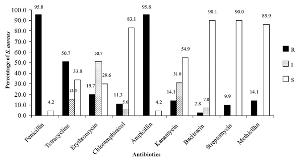
The antibiotic resistance profiles of Staphylococcus aureus isolates revealed varying levels of resistance across the antibiotic tested (Figure 1). The highest resistance rates were observed for penicillin and ampicillin, with 95.8% of isolates classified as resistant to each antibiotic. Moderate resistance was also detected against tetracycline (50.7%) and erythromycin (19.7%), while 50.7% of isolates showed intermediate susceptibility to erythromycin. In contrast, resistance to chloramphenicol (11.3%), kanamycin (14.1%), bacitracin (2.8%), streptomycin (9.9%), and methicillin (14.1%) was low among the isolates tested. Intermediate susceptibility was most frequently observed for kanamycin (31.0%) and bacitracin (7.0%). These findings indicate that resistance was predominantly associated with β-lactam antibiotics, particularly penicillin and ampicillin, whereas lower resistance levels were observed for the remaining antimicrobial agents. Their multiple antibiotic resistance (MAR) index values ranged from 0 to 0.88 (Table 1). The reference strain (S. aureus TISTR 1466) showed a MAR index of 0. The majority of clinical isolates exhibited MAR indices of 0.22 (37.50%) and 0.33 (34.72%). Higher MAR indices were less frequent, with 8.33% of isolates showing a MAR index of 0.44, and smaller proportions exhibiting indices of 0.55, 0.66, 0.77, and 0.88.
As shown in Table 1, MHMR index values ranged from 0.57 to 0.71, thus indicating that all isolates were resistant to more than half of the heavy metals tested. A minority of isolates (12.50%) exhibited an MHMR index of 0.57, while the majority (87.50%) displayed an index of 0.71. Resistance to individual heavy metals across concentrations ranging from 0.1 to 80.0 mM is shown in Table 2. At 0.1 mM, all isolates (100%) were resistant to silver (Ag), chromium (Cr), lead (Pb), and zinc (Zn), with high resistance also observed for copper (Cu; 88.1%), mercury (Hg; 88.5%), and cobalt (Co; 84.2%). Chromium exhibited the highest tolerance, with all isolates resistant up to 10.0 mM and nearly complete resistance maintained at 20.0–40.0 mM. Lead resistance persisted up to 10.0 mM but declined sharply thereafter. Resistance to Ag, Co, Cu, Hg, and Zn was limited to lower concentrations, with no resistance observed at higher levels.
|
[Metals] (mM) |
0.1 |
0.5 |
1.0 |
5.0 |
10.0 |
20.0 |
40.0 |
80.0 |
|
Ag |
100.0 |
82.9 |
0.0 |
0.0 |
0.0 |
0.0 |
0.0 |
0.0 |
|
Co |
84.2 |
6.5 |
0.0 |
0.0 |
0.0 |
0.0 |
0.0 |
0.0 |
|
Cr |
100.0 |
100.0 |
100.0 |
100.0 |
100.0 |
98.6 |
98.6 |
0.0 |
|
Cu |
88.1 |
15.7 |
6.5 |
1.3 |
0.0 |
0.0 |
0.0 |
0.0 |
|
Hg |
88.5 |
6.5 |
0.0 |
0.0 |
0.0 |
0.0 |
0.0 |
0.0 |
|
Pb |
100.0 |
100.0 |
100.0 |
100.0 |
100.0 |
0.0 |
0.0 |
0.0 |
|
Zn |
100.0 |
100.0 |
100.0 |
14.4 |
0.0 |
0.0 |
0.0 |
0.0 |
Phenotypic screening assay identified 13 resistance detections, corresponding to 12 unique resistant S. aureus isolates, comprising 9 MRSA, 3 VISA, and 1 VRSA (Table 1). One isolate (CRP41) was classified as both MRSA and VRSA. Among MRSA isolates, six were recovered from Hospital A, two from Hospital B, and one from Hospital C. All VISA and VRSA isolates were detected exclusively from Hospital A. The resistant isolates exhibited MAR indices ranging from 0.22 to 0.88. Isolates CRP44, CRP45, and CRP46 showed the highest MAR index (0.88), with resistance to eight antibiotics. Moderate MAR values (0.55–0.77) were observed in isolates CRP35, CRO01, CRO10, and CRK01, whereas the lowest MAR index (0.22) occurred in isolates CRP41 and CRP47. Notably, bacitracin (10 U) was the only antibiotic that remained effective against all resistant S. aureus isolates, including those with the highest MAR indices. All MRSA isolates demonstrated resistance to multiple heavy metals, with MHMR indices ranging from 0.57 to 0.71. Most were resistant to Cu, Cr, Hg, Pb, and Zn, although the absence of mercury resistance in some isolates resulted in lower MHMR values. VISA isolates exhibited lower MAR indices (0.22–0.33), primarily resistant to ampicillin, penicillin-G, and tetracycline, but all showed high heavy metal resistance (MHMR = 0.71). The VRSA isolate also displayed a low MAR index (0.22) but resistance to all tested heavy metals.
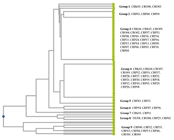
Cluster analysis based on combined antibiotic and heavy metal resistance profiles grouped the 71 S. aureus isolates into nine major clusters (Figure 2). Group 9 comprised nine isolates with high MAR indices (0.55–0.88), including six MRSA isolates. Groups 3 and 4 showed moderate resistance (MAR = 0.22–0.33), with Group 3 including CRP41 (MRSA/VRSA) and CRP57 (VISA), and Group 4 consisting of two VISA isolates (CRP22 and CRP51). Group 8 contained three relatively susceptible isolates (MAR = 0.11) and the fully susceptible type strain (MAR = 0.00).
This study provides a comprehensive phenotypic assessment of antibiotic and heavy metal resistance among Staphylococcus aureus isolates recovered from hospitals. The findings reveal a high prevalence of resistance to commonly used antibiotics, widespread tolerance to multiple heavy metals, and distinct clustering of isolates based on combined resistance profiles. Collectively, these results underscore the role of sustained selective pressure in shaping resistance patterns within hospital-associated S. aureus populations.
The extremely high resistance rates to penicillin and ampicillin observed in this study are consistent with global reports documenting widespread β-lactam resistance in S. aureus [28,29]. This resistance is primarily attributed to the production of β-lactamase enzymes and alterations in penicillin-binding proteins, which reduce antibiotic efficacy [30]. The notable resistance to tetracycline and erythromycin further reflects the extensive and prolonged use of these antibiotics in clinical settings, which promotes the selection and persistence of resistant strains. In contrast, low resistance to chloramphenicol, bacitracin, streptomycin, kanamycin, and methicillin indicates that these antibiotics remain effective against the majority of isolates examined. Nevertheless, the detection of methicillin resistance in a subset of isolates is clinically significant, as MRSA infections are associated with limited treatment options and an increased morbidity [31].
Analysis of the MAR index further supports these observations, with most clinical isolates exhibiting MAR index values >0.2, suggesting exposure to environments with frequent antibiotic use and possible selective pressure [23]. These elevated MAR values suggest strong and sustained selective pressure within hospital settings, where antimicrobial agents are routinely and intensively used [32]. The complete susceptibility of the S. aureus type strain (TISTR 1466), reflected by a MAR index of 0.00, confirms its appropriateness as a reference strain and highlights the acquired nature of resistance among clinical isolates. The MAR index observed in this study reflects a relatively high proportion of S. aureus isolates associated with environments subjected to sustained antimicrobial exposure. However, the MAR index is a cumulative measure and does not consider overlap among antibiotics within the same antimicrobial classes. In this study, several tested antibiotics, particularly β-lactams and aminoglycosides, share related mechanisms of action or resistance pathways. As a result, resistance to multiple agents within the same class may contribute to higher MAR values without necessarily representing independent resistance events. Despite this limitation, the MAR index remains a useful epidemiological indicator for assessing overall antimicrobial exposure pressure and identifying high-risk contamination sources in hospital settings. Accordingly, MAR values in this study were interpreted as a general measure of resistance burden rather than a mechanism-specific descriptor. Multidrug resistance (MDR), as defined by Magiorakos et al. [22], provides a more standardized classification based on resistance across antimicrobial categories. Although MDR analysis was not included as a primary endpoint in this study, the available isolate-level susceptibility data would allow such classification in future analyses. Together, MAR and MDR approaches may provide complementary perspectives, where MAR reflects overall exposure pressure and MDR offers standardized resistance categorization.
Apart from antibiotic resistance, this study demonstrates a high prevalence of resistance to multiple heavy metals among S. aureus isolates. All isolates exhibited MHMR index values exceeding 0.50, thereby indicating resistance to more than half of the tested metals. The predominance of isolates with an MHMR index of 0.71 suggests prolonged and repeated exposure to heavy metals in the environments from which these isolates were recovered [33,34]. Notably, tolerance to chromium and lead was observed across a wide range of concentrations. Both metals are environmentally persistent and are commonly associated with industrial effluents and environmental contamination. Their presence in surrounding environments, including areas impacted by hospital waste streams, may contribute to selective pressure on microbial populations [35]. In contrast, resistance to silver, copper, mercury, cobalt, and zinc was largely confined to lower concentrations, reflecting their higher toxicity and the limited adaptive capacity of S. aureus under elevated metal stress [36].
The concurrent occurrence of elevated MAR and MHMR indices among the S. aureus isolates suggests a potential linkage between antibiotic and heavy metal resistance. Resistance determinants for both antibiotics and heavy metals are often co-localized on mobile genetic elements, including plasmids, transposons, and integrons [13]. Consequently, exposure to heavy metals may indirectly select for antibiotic resistance even in the absence of antibiotic pressure – which is a phenomenon known as co-selection [37]. Hospital environments, where antimicrobial agents and certain metal-based materials (e.g., silver- or copper-containing surfaces and coatings) are used, may serve as hotspots for the persistence and dissemination of resistant bacterial strains.
Cluster analysis based on combined antibiotic and heavy metal resistance profiles revealed substantial phenotypic diversity among the S. aureus isolates while also identifying distinct resistance-associated clusters. The presence of clusters characterized by high MAR index values suggests the circulation of predominant lineages with enhanced resistance potential. Conversely, clusters containing isolates with lower MAR values, including the type strain, indicate the coexistence of less resistant or susceptible populations within the same hospital environments. These clustering patterns support the notion that resistance traits are not randomly distributed but are instead associated with specific phenotypic lineages shaped by shared selective pressures [38]. Such patterns may reflect common sources of contamination, transmission routes, or similar environmental exposures within hospital settings, emphasizing the importance of integrated surveillance and targeted infection control strategies [39].
This study has several limitations that should be acknowledged. First, the assessment of antibiotic and heavy metal resistance was based solely on phenotypic methods. While these approaches provide reliable information on resistance patterns, they do not allow identification of the specific genetic determinants or mechanisms responsible for resistance. The lack of molecular analyses, such as the detection of resistance genes or characterization of mobile genetic elements, limits the ability to confirm genetic linkage or co-localization of antibiotic and heavy metal resistance traits. Second, isolates were obtained from a limited number of hospitals within a single geographic area. Differences in antibiotic usage, infection control practices, and environmental conditions among healthcare facilities may influence resistance profiles; therefore, the findings may not be generalizable to other hospital settings. In addition, the unequal distribution of isolates among the hospitals may have affected comparisons of resistance prevalence. Third, heavy metal resistance was evaluated using fixed threshold concentrations under laboratory conditions. These concentrations may fail to accurately represent the variable and often lower levels of heavy metals present in hospital environments. Furthermore, possible interactions among different heavy metals or between heavy metals and disinfectants were not examined and may influence resistance expression in situ. Finally, this investigation was cross-sectional in nature. As a result, changes in resistance patterns over time could not be assessed. Long-term surveillance would be necessary to evaluate trends in antibiotic and heavy metal resistance and to determine the persistence or emergence of resistant S. aureus populations in hospital environments.
This study demonstrates a high prevalence of antibiotic resistance and widespread tolerance to multiple heavy metals among S. aureus isolates recovered from hospitals. Resistance to β-lactam antibiotics was particularly common, and most isolates exhibited multiple antibiotic resistance, indicating strong selective pressure within healthcare settings. In addition, all isolates showed resistance to several heavy metals, with especially high tolerance to chromium and lead. The occurrence of MRSA, VISA, and VRSA isolates, together with elevated MAR and MHMR indices, highlights the persistence of resistant S. aureus populations in hospitals. Cluster analysis further revealed distinct resistance-associated groups, suggesting the circulation of phenotypically related strains shaped by shared environmental pressures. These findings emphasize the need for continued surveillance of both antibiotic and heavy metal resistance and support the implementation of integrated infection control and environmental management strategies to limit the spread of resistant S. aureus in healthcare facilities.
We would like to thank Mae Fah Luang University for funding this work.
In loving memory of the corresponding author’s beloved mother, Boontiwa Chukeatirote (3 April 1940 – 13 October 2025), whose enduring love, kindness, and wisdom continue to guide and inspire his life and work across the past, present, and future.
No competing financial interest or personal relationship could have appeared to influence the work reported in this paper.
N. S.: investigation, writing – original draft preparation.
E. C.: conceptualization, methodology, supervision, formal analysis, writing – review and editing.