![[The figure shows three brain MRI views. The left and middle images are axial susceptibility-weighted MRI sequences, and the right image is a coronal T1-weighted post-contrast image. An arrow labeled ‘A’ marks an elongated focus in the region of the left central sulcus, described as hemosiderin microdeposition with microcalcification, best visualized on the SWAN calcium filter image. The same finding is not clearly visible on the other MRI sequences. An arrow labeled ‘B’ indicates the nearby Trolard vein, which follows its usual course and shows no visible abnormality. There are no visible perifocal cortical changes suggesting cytotoxic edema or encephalomalacia.]](https://www.zurnalai.vu.lt/AML/article/download/45426/version/41570/42954/140907/Kaybryte-1_pav.jpg)
Acta medica Lituanica ISSN 1392-0138 eISSN 2029-4174
2026. Vol. 33. No 2. Online ahead of print DOI: https://doi.org/10.15388/Amed.2026.33.2.5
Gertrūda Kaubrytė*
Vilnius University, Faculty of Medicine, Vilnius, Lithuania
E-mail: gertruda.kaubryte@mf.stud.vu.lt
ORCID ID https://orcid.org/0009-0009-2880-483X
Rūta Samaitienė-Aleknienė
Pediatrics Centre, Clinic of Children’s Diseases; Faculty of Medicine, Vilnius University, Vilnius, Lithuania
E-mail: ruta.samaitiene@santa.lt
ORCID ID https://orcid.org/0000-0001-6503-1578
Abstract. Background: Despite the difficulties of diagnosing intracranial hypotension (IH), the literature focusing on specific IH misdiagnoses remains scarce. We report on the case of a pediatric patient with spontaneous intracranial hypotension (SIH) caused by thecal sac dilatation due to Marfan syndrome, who was initially misdiagnosed. Additionally, a systematic literature review was conducted, focusing specifically on misdiagnoses of IH.
Materials and Methods: The material search for the literature review was performed across PubMed, Scopus, and Web of Science databases. The inclusion criteria were: published within the last ten years, involved human participants, written in English, and inclusion of case reports or series with a clear diagnostic pathway, in which, a specified initial misdiagnosis was followed by IH as the final diagnosis. Publications were excluded if they did not meet these criteria. Due to the nature of the case reports, reporting bias and certainty assessment were not formally evaluated. Additionally, a single-patient case report was retrospectively analyzed.
Results: A 13-year-old woman presented to the emergency department complaining of a severe postural headache, nausea, sleepiness, generalized weakness, dizziness, and loss of appetite. An asymptomatic venous thrombosis and functional headaches were suspected. The patient was transferred to a third-level hospital, where a magnetic resonance imaging scan of the whole spine revealed thecal sac dilatation. SIH was diagnosed, and, after 3 weeks of conservative treatment, the patient was discharged from the hospital with full recovery. In the systematic review, 24 studies containing 28 patients were included in the final report. The most frequent initial misdiagnoses were Chiari I malformation (39.29%) and migraine (21.43%). 75% of studies identified SIH as a cause of IH, 16.67% determined it was iatrogenic, 4.17% – traumatic, and 1 case report did not include the cause of IH.
Conclusions: While the case report illustrates the difficulties in diagnosing IH, the systematic review distinguishes initial misdiagnoses of this condition, of which, the most common are migraine and Chiari I malformation.
Keywords: intracranial hypotension, Marfan syndrome, systematic review, case report, misdiagnosis.
Santrauka. Įvadas: Nors intrakranijinė hipotenzija (IH) yra liga, kurią sudėtinga diagnozuoti, literatūros, kurioje rašoma apie specifines IH misdiagnozes, mažoka. Pristatome spontaninės intrakranijinės hipotenzijos (SIH) dėl Marfano sindromo sukelto jungiamojo audinio trapumo pediatrinio paciento klinikį atvejį, kurį vertinant iš pradžių buvo suformuluota neteisinga diagnozė. Sisteminė literatūros, kurioje koncentruotasi į IH misdiagnozes, apžvalga buvo atlikta.
Metodai: Literatūros ieškota „PubMed“, „Scopus“ ir „Web of Science“ duomenų bazėse. Įtraukimo kriterijai buvo šie: publikuota per paskutinius dešimt metų, tyrime dalyvavo žmonės, parašyta anglų kalba, publikacijoje aprašomi klinikiniai atvejai ar jų serijos, jose nurodomas aiškus diagnostinis kelias, kuriame išskirta IH specifinė misdiagnozė. Publikacijos, neatitikusios įtraukimo kriterijų, buvo atmestos. Kadangi buvo renkama klinikinių atvejų aprašymų informacija, publikavimo šališkumas ir įrodymų patikimumas nebuvo vertinti. Be to, vieno paciento klinikiniai duomenys analizuoti retrospektyviai.
Rezultatai: 13 metų pacientė atvyko į priėmimo skyrių dėl stipraus ortostatinio galvos skausmo, pykinimo, mieguistumo, bendro silpnumo, galvos svaigimo ir apetito nebuvimo. Pirminė diagnozė – asimptominė venų trombozė ir funkcinis galvos skaumas. Pacientė buvo perkelta į trečio lygio ligoninę, joje atliktas viso stuburo magnetinio rezonanso vaizdinis tyrimas parodė duralinio maišo išsiplėtimą. Diagnozuota SIH ir po trijų savaičių konservatyvaus gydymo pacientė išleista namo be liekamųjų reiškinių. Į sisteminę apžvalgą įtrauktos 24 publikacijos, kuriose buvo aprašyti 28 klinikiniai atvejai. Nustatytos dažniausios IH misdiagnozės buvo Chiari I malformacija (39,29 %) ir migrena (21,43 %). Dauguma (75 %) studijų nurodė, kad IH buvo spontaninė, 16,67 % – jatrogeninė, 4,17 % – trauminė, vieno atvejo aprašyme IH priežastis nenurodyta.
Išvados: Pristatytas atvejis iliustruoja sunkumus diagnozuojant IH, o sisteminė apžvalga leidžia išskirti konkrečias IH misdiagnozes, iš kurių Chiari I malformacija ir migrena yra pačios dažniausios.
Raktažodžiai: intrakranijinė hipotenzija, Marfano sindromas, sisteminė apžvalga, atvejo aprašymas, misdiagnozė.
___________
* Corresponding author
Received: 17/02/2026. Revised: 13/04/2026. Accepted: 15/04/2026
Copyright © 2026 Gertrūda Kaubrytė, Rūta Samaitienė-Aleknienė. Published by Vilnius University Press.This is an Open Access article distributed under the terms of the Creative Commons Attribution License, which permits unrestricted use, distribution, and reproduction in any medium, provided the original author and source are credited.
Intracranial hypotension (IH) is defined by Cerebrospinal Fluid (CSF) pressure decrease less than 60 mm H2O, with postural headache being the hallmark symptom [1]. Headache attributed to low CSF pressure is usually accompanied by neck pain, tinnitus, hearing changes, photophobia, and nausea, and it resolves after normalization of CSF pressure or management of CSF leak. It is diagnosed by the following diagnostic criteria: low CSF pressure and/or evidence of CSF leakage on imaging, and headache has developed in temporal relation to these diagnostic findings, or led to its discovery, and it cannot be better accounted for by another ICHD-3 (International Classification of Headache Disorders) diagnosis [2]. The ICHD-3 distinguishes three types of headache attributed to low CSF pressure: 1) Post-dural puncture headache, which is diagnosed when headache has developed within 5 days of the dural puncture [3]; 2) CSF fistula headache which is diagnosed when a procedure has been performed, or trauma has occurred, which can cause CSF fistula, and headache has developed in temporal relation to the procedure or trauma [4]; 3) Headache attributed to Spontaneous Intracranial Hypotension (SIH) which is diagnosed when there is absence of a procedure or trauma known to be able to cause CSF leakage, and headache has developed in temporal relation to occurrence of low CSF pressure or CSF leakage, or has led to its discovery [5]. As of now, the medical community lacks comprehensive data covering all IH types due to some types being dependent on medical procedures or traumatic events; therefore, we present the statistics of SIH. It most commonly affects middle-aged women, whereas the overall incidence of this condition is approximately 5 in 100000 [1]. Although it is not a rare condition, IH, and especially SIH, is frequently misdiagnosed. A cross-sectional online survey conducted in 2022 found that, on average, patients who experienced SIH symptoms had three appointments with their general practitioner before being referred to a specialist, and just over a half were correctly diagnosed by the first specialist they saw [6]. This means unnecessary treatments and diagnostic measurements, and, most importantly, delayed treatment, which can lead to serious complications such as cerebral venous thrombosis or posterior circulation infarction [1]. Despite this issue, the literature focusing on specific IH misdiagnoses remains scarce, with a lack of systematic literature reviews. There is a reason to discuss the most common misdiagnoses of IH and present more case reports with interesting diagnostic pathways. Herein, we report on the case of a 13-year-old patient with SIH caused by thecal sac dilatation due to Marfan syndrome. This case offers several unique aspects: 1) the young age of the patient; 2) pre-existing genetic syndrome; 3) conservative treatment plan; and 4) initial misdiagnosis. Additionally, we have conducted a systematic literature review, which, to the best of our knowledge, is the first of this type of publication, focusing specifically on misdiagnoses of intracranial hypotension.
The case report was written following CARE guidelines (see Supplementary File No. 1). An informed consent form was signed by the patient and the patient’s mother.
The process of systematic review followed the Preferred Reporting Items for Systematic Reviews and Meta-Analysis (PRISMA) 2020 guidelines (see Supplementary File No. 2). The review protocol has not been prepared and registered.
The material search for the literature review was performed across PubMed, Scopus, and Web of Science databases on 25 November 2025. A 10-year publication date, case reports, full-text, and English-language filters were applied wherever available. Search strategies were as follows: “intracranial hypotension”[Mesh] OR “intracranial hypotension”[tiab] OR “spontaneous intracranial hypotension”[tiab] OR “SIH”[tiab] OR “CSF leak”[tiab] OR “cerebrospinal fluid leak”[tiab] OR “spinal CSF leak”[tiab] OR “CSF hypovolemia”[tiab] (PubMed); TITLE-ABS-KEY( “intracranial hypotension” OR “spontaneous intracranial hypotension” OR “SIH” OR “CSF leak” OR “cerebrospinal fluid leak” OR “spinal CSF leak” OR “CSF hypovolemia”) AND TITLE-ABS-KEY(“case report” OR “case reports” OR “case series”) (Scopus); TOPIC: (“intracranial hypotension” OR “CSF leak” OR “spontaneous intracranial hypotension”) AND (“case report” OR “case series”) (Web of Science).
The literature review only included publications that 1) were published within the last ten years; 2) involved human participants; 3) were written in English; 4) included case reports or case series with a clear diagnostic pathway, in which a specified initial misdiagnosis was followed by intracranial hypotension as the final diagnosis. Publications were excluded if they did not meet the inclusion criteria.
Relevant studies were then imported into the Zotero reference management software. After evaluating titles and abstracts, a human reviewer selected certain publications; no automatic tools were used. Subsequently, the full texts selected were assessed for inclusion or exclusion in accordance with predefined criteria in the Zotero reference management software.
To manually extract data from the publications included in the literature review, a template was developed in Microsoft Excel. It determined the first author, the year of the publication, the age and sex of the patients, and the initial misdiagnosis of intracranial hypotension. Missing or unclear information was recorded as ‘not reported’, and no assumptions were made. One reviewer assessed the quality of case reports in accordance with the CARE guidelines.
Due to the heterogeneity of the case reports included in the literature review, narrative synthesis was implemented in preference to meta-analysis. Possible explanations for heterogeneity were not explored. Extracted data were systematized by using descriptive statistics. The results are presented in the form of texts and tables.
Case reports are known to be biased, but, due to the nature of this publication type, reporting bias was not formally evaluated. Certainty assessment was not executed, as no quantitative synthesis was made. Case reports inherently represent low-level evidence.
A 13-year-old woman presented to the emergency department complaining of a severe headache (mostly in the frontal area), nausea, sleepiness, generalized weakness, dizziness, and loss of appetite. While standing or sitting, headache severity was 10 according to the Visual Analogue Scale (VAS), accompanied with nausea and dizziness. While in a supine position, headache severity decreased to 3 according to the VAS, and nausea and dizziness also improved. The patient was afebrile, with no signs of head trauma or infection. She had been diagnosed with Marfan syndrome, has an aortic aneurysm, atrial septal defect, hypoplastic left heart syndrome, and both eye lenses are implanted because of aphakia. The patient’s mother and sister had also been diagnosed with Marfan syndrome. Five years earlier, the patient had been hospitalized due to acute headaches and vomiting. Then, the head MRI (Magnetic Resonance Imaging) scan showed superior sagittal sinus thrombosis, and 0.3 ml of low-molecular-weight heparin (LMWH) two times per day was prescribed subcutaneously. The patient then made a complete recovery.
During physical examination, the patient exhibited several Marfanoid features, including a tall, thin stature, disproportionately long arms and legs, dolichocephaly, malar hypoplasia, and retrognathia. The patient was underweight with a BMI (Body Mass Index) of 12.87. The neurological examination showed bilateral foot clonus, a slight reflex asymmetry, and hyperreflexia in the legs.
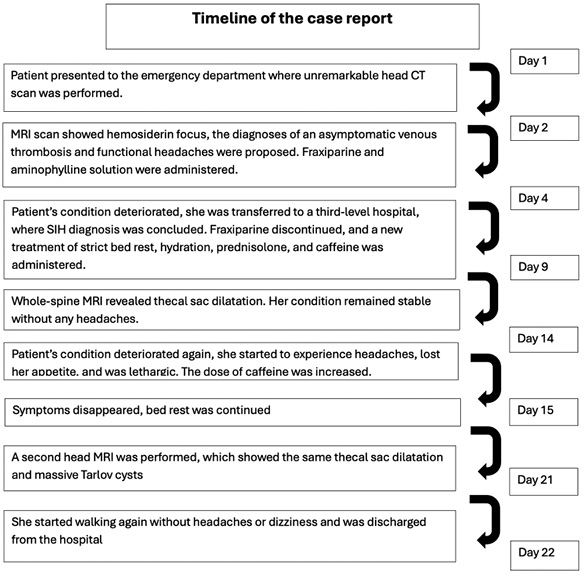
On the admission day, the head CT (Computer Tomography) scan was unremarkable. The next day, the head MRI scan showed a small, elongated hemosiderin focus in the left central sulcus region and an asymmetrically narrow Trolard anastomotic vein adjacent to it (Figure 1). An asymptomatic venous thrombosis and functional headaches were suspected. Urine and blood analyses showed slightly decreased SPA (52%) and PO2 (64.4 mmHg). Due to the risk of possible venous thrombosis, 0.6 ml of LMWH twice daily was administered subcutaneously. Additionally, 0.5 mg/kg/h of aminophylline solution and intravenous fluids were administered. After four hospitalization days, the patient’s condition worsened, and IH was suspected. Because of limited medical examination possibilities, the patient was transported to a third-level hospital. There, the head MRI scan was reevaluated by a radiologist: the venous thrombosis diagnosis was rejected; therefore, LMWH was discontinued. A new diagnosis of spontaneous intracranial hypotension associated with Marfan syndrome was formulated. The following treatment was prescribed: strict bed rest, hydration, 1mg/kg/d of prednisolone for five days, and caffeine. On the ninth hospitalization day, a whole-spine MRI scan was performed, which revealed thecal sac dilatation (57 x 22 mm) with expansion into the foraminal openings and Tarlov cysts. Additionally, fluid was present in the pelvis (Figure 2). This test confirmed the hypothesis for SIH; the treatment was continued, and her condition remained stable, with no headaches for five days. On the fourteenth hospitalization day, the patient started to experience headaches again (VAS up to 5 points), lost her appetite, and was lethargic. The dose of caffeine was increased to the equivalent of six cups of coffee. The symptoms disappeared the next day, and bed rest was continued. After three weeks of hospitalization, a second head MRI scan was performed, which showed the same thecal sac dilatation and massive Tarlov cysts. There was no CSF leakage. After 21 days of conservative treatment, she started walking again without headaches or dizziness, and was discharged from the hospital on day 22 (Figure 3).
![[Three T2-weighted spine MRI images are shown from left to right: sagittal, axial Cube, and 3D FIESTA sequences. Together, the images demonstrate dural ectasia, with ballooning and widening of the dural sac in the thoracic, lumbar, and sacral regions of the spine. In the sagittal image on the left, the dural sac appears enlarged along the lower spine. In the axial and 3D FIESTA images, multiple perineural cysts, also known as Tarlov cysts, are visible in the sacral region. Some of these cysts extend distally from the sacrum along the course of the nerve roots. Label ‘A’ marks the sacral perineural cystic extensions.]](https://www.zurnalai.vu.lt/AML/article/download/45426/version/41570/42954/140904/Kaybryte-2_pav.jpg)
Although the conservative treatment was successful and the patient left the hospital being asymptomatic, there were several adverse events related to the prolonged strict bed regime: obstipation, a disrupted sleep cycle, mood swings, and skipping school. During the first week, while being able to stand, the patient experienced leg and back pain, as well as fatigue. During the follow-up two weeks later, the patient stated she was better and was not experiencing any headaches; she was also temporarily homeschooling and planning to return to school next week. During a second follow-up, 3 months after the hospital discharge, the patient denied recurrence of postural headaches and attended school as usual.
A total of 2290 publications were initially identified through database searches. 1119 records were removed before screening: 1114 duplicates, 2 marked as ineligible by automation tools, 2 errata were removed for not containing original case data, and 1 record was removed because it only contained corrections of a previous publication. After screening the titles and abstracts of 1171 publications, 425 records were determined eligible to retrieve. 41 records were failed to retrieve, and 384 full-text articles were assessed for eligibility. Of these, 24 records met the criteria and were included in the final report (Figure 3).
![[The figure is a PRISMA-style flow diagram titled “Identification of studies via databases and registers”. It is organized into three stages: Identification, Screening, and Included.
In the identification stage, records were identified from three databases. Before screening, part of records were removed for the following reasons: 1,114 duplicate records, 2 records marked as ineligible by automation tools, and 3 records removed for other reasons. In the screening stage, 1,171 records were screened. Of these, 746 records were excluded. After screening, 425 reports were sought for retrieval. Of these, 41 reports could not be retrieved. A total of 384 reports were assessed for eligibility. Reports were excluded for the following reasons: the initial diagnosis was correct in 224 reports, no intracranial hypotension diagnosis was presented in 63 reports, the wrong initial diagnosis was unspecified in 19 reports, no clear diagnostic pathway was described in 45 reports, and the article was not in English in 9 reports. In the ‘Included’ stage, 24 studies were included in the review.]](https://www.zurnalai.vu.lt/AML/article/download/45426/version/41570/42954/140906/Kaybryte-4_pav.jpg)
The studies included in this case report were published between 2015 and 2025. Out of 28 (one study contains 5 included cases) case reports, 17 (60.71%) patients were females, and the mean age of the patients was 40.43 years. Initial misdiagnoses of these reports included neoplasm [7], migraine [8–13], Chiari I malformation [11,14–19], muscle strain [12], occipital neuralgia [12], tension headaches [12], temporomandibular joint disorder [12], new daily persistent headache [12], impending transtentorial herniation [13], sinus headache [20], meningitis [21], intracranial hypertension [22,23], posttraumatic headache [24], cervicogenic headache [25], Ménière’s disease [26], head injury [27], musculoskeletal cervicalgia [28], heartburn [29], viral meningitis [30]. Among 24 studies, 18 (75%) identified SIH as a cause of IH, 4 (16.67%) determined it was iatrogenic, 1 (4.17%) assessed it was traumatic, and 1 case report did not include the cause of intracranial hypotension (Table 1).
|
|
First |
Publication year |
Reporting patient’s |
Reporting patient’s age |
Initial |
Cause |
|
1 |
Bray et al. |
2016 |
male |
58 |
Neoplasm |
Spontaneous IH |
|
2 |
Daripa et al. |
2022 |
female |
32 |
Migraine |
Spontaneous IH |
|
3 |
Lee et al. |
2018 |
female |
43 |
Migraine |
Spontaneous IH |
|
4 |
Rettenmaier et al. |
2017 |
female |
62 |
Migraine |
Spontaneous IH |
|
5 |
Agresta et. al |
2020 |
female |
32 |
Migraine, Chiari I malformation |
Spontaneous IH |
|
6 |
Ruggeri-McKinley et al. |
2016 |
female |
28 |
Muscle strain, exercise induced migraines, occipital neuralgia, tension headaches, temporomandibular joint disorder, new daily persistent headache |
Spontaneous IH |
|
7 |
Inamasu et al. |
2015 |
female |
58 |
Migraine, impending transtentorial herniation |
Spontaneous IH |
|
8 |
Nisson et al. |
2021 |
female |
2 |
Chiari I malformation |
Spontaneous IH |
|
9 |
Chan et al. |
2021 |
4 females, 1 male |
25, 26, 57, 29, 41 |
Chiari I malformation |
Spontaneous IH |
|
10 |
Huls et al. |
2022 |
male |
50 |
Chiari I malformation |
Spontaneous IH |
|
11 |
Kingston et al. |
2017 |
male |
44 |
Chiari I malformation |
Spontaneous IH |
|
12 |
Mostofi et al. |
2016 |
male |
46 |
Chiari I malformation |
Spontaneous IH |
|
13 |
Park et al. |
2023 |
female |
51 |
Chiari I malformation |
Spontaneous IH |
|
14 |
Redon et l. |
2020 |
male |
35 |
Sinus headache |
Spontaneous IH |
|
15 |
Wosaibai et al. |
2020 |
male |
8 |
Meningitis |
Iatrogenic (after lumbar puncture) |
|
16 |
O’Neill et al. |
2021 |
female |
47 |
Intracranial hypertension |
Iatrogenic (after thoracic laminectomy) |
|
17 |
Subramaniam et al. |
2021 |
male |
50 |
Intracranial hypertension |
Spontaneous IH |
|
18 |
Richard et al. |
2016 |
male |
45 |
Posttraumatic headache |
Traumatic IH |
|
19 |
Primalani et al. |
2019 |
male |
49 |
Cervicogenic headache |
Spontaneous IH |
|
20 |
Cahal et al. |
2023 |
female |
39 |
Ménière’s disease |
Spontaneous IH |
|
21 |
Federspiel et al. |
2020 |
female |
56 |
Head injury |
Iatrogenic (after thoracotomy) |
|
22 |
Bueno et al. |
2025 |
female |
40 |
Musculoskeletal cervicalgia |
Iatrogenic (after epidural anesthesia) |
|
23 |
Bortolato et al. |
2020 |
male |
45 |
Heartburn |
Spontaneous IH |
|
24 |
Qureshi et al. |
2018 |
female |
34 |
Viral meningitis |
no data |
The completeness of the studies included in this report was assessed in accordance with the CARE guidelines. Part of the reports failed to include the type of their publication type in the title, some studies did not list the key words, several abstracts were incomplete, and a single report did not have a complete introduction and discussion. All the publications included de-identified patient information, primary concerns and symptoms of the patient, clinical findings, diagnostic methods and diagnosis, and none of them contained a patient’s perspective. Almost all included detailed diagnostic assessment and therapeutic intervention sections. One report assessed the limitations of their publication. Publication bias is likely to occur due to cases being unusual or severe and the reluctance to address the negative aspects of the case reports, such as initial misdiagnoses. Overall certainty of evidence is low.
While the case presented has a unique and detailed SIH diagnostic pathway and shows successful conservative treatment for this condition, it has several limitations, including single-patient observation, limited diagnostic measurements, and single-center experience. The presented systematic literature review was written following the PRISMA guidelines and offers a broad overview of the existing literature; however, it comes with limitations such as selective reporting, which inherently represents low-level evidence, heterogeneity of clinical presentation, and underreporting of misdiagnoses. Additionally, the review protocol has not been prepared and registered, and the literature was reviewed by a single person.
Although the presented case had a favorable outcome, it highlights several difficulties when diagnosing SIH. We hypothesize that previous superior sagittal sinus thrombosis, which occurred 5 years before this hospitalization, was also caused by SIH. Therefore, the accurate diagnosis could have been delayed for 5 years. Additionally, diagnostic setbacks occurred in the hospitalization of this case report, too, and an unjustified diagnosis of asymptomatic venous thrombosis and functional headache was made. The formulation of the first diagnosis was challenging due to the lack of diagnostic measurements, the rare condition, and limited information about the patient’s clinical course. Additionally, this situation can be explained by the anchoring effect, which was introduced in 1974 by Tversky and Kahneman. It describes how the initially presented value can influence the decision-maker’s bias in favor of that value [31]. The decision to diagnose asymptomatic venous thrombosis could have been driven by the previous diagnosis of sinus thrombosis, and it may have resulted in missing the new information, e.g., the postural feature of the headache, which could have led to the correct diagnosis. Knowing all the obstacles in diagnosing this case, the decision to transfer the patient to a third-level hospital, where the involvement of a multidisciplinary team (children’s neurologist, children’s neurosurgeon, and radiologist) ensured more comprehensive care, was reasonable. Overall, the patient’s treatment in this case was successful with no residual deficits, and therefore this case report acts as a valuable lesson to medical practitioners while allowing full recovery.
Just as the case presented shows, IH is usually misdiagnosed. The literature analysis shows that the most common misdiagnoses are Chiari I malformation and migraine. According to our literature review, 60.71% of misdiagnosed patients were females, but it is known that IH affects more female patients than males. The largest number of initial misdiagnoses occurs in cases with SIH, but our review suggests that iatrogenic and even traumatic IH are also misdiagnosed. A retrospective clinical study published in 2003 listed misdiagnoses of SIH; these included migraine (33%), meningitis (18%), psychogenic/malingering (12%), tension headache (6%), subarachnoid hemorrhage (6%), Chiari malformation (6%), posterior cervical strain (3%), ocular myasthenia gravis (3%), transient ischemic attack (3%), benign exertional headache (3%), subdural hematoma (3%), cervical radiculopathy (3%) [32]. These findings partially match the results of our systematic literature review: we have found a greater misdiagnosis rate of Chiari I malformation (39.29%), similar incidence of migraine (21.43%) and muscle strain (3.57%) misdiagnoses, lower rate of meningitis (3.57%) and tension headache (3.57%) inaccurate initial diagnoses. Another report included different initial diagnoses such as psychogenic/malingering headache, subarachnoid hemorrhage, ocular myasthenia gravis, transient ischemic attack, benign exertional headache, subdural hematoma, and cervical radiculopathy [32]. In addition, we report the following misdiagnoses which were not included in this study: neoplasm (3.57%), occipital neuralgia (3.57%), temporomandibular joint disorder (3.57%), new daily persistent headache (3.57%), impending transtentorial hernation (3.57%), sinus headache (3.57%), intracranial hypertension (7.14%), posttraumatic headache (3.57%), cervicogenic headache (3.57%), Ménière’s disease (3.57%), head injury (3.57%), musculoskeletal cervicalgia (3.57%), and heartburn (3.57%). The patient in our case report and many others received unnecessary treatment, diagnostic measures, and received delayed appropriate treatment because of the initial diagnosis being wrong. Consequently, it is beneficial to discuss several of the presently mentioned misdiagnoses that have been misdiagnosed more frequently.
Migraine is a type of primary headache disorder that has recurring attacks which usually last 4–72 hours, and is accompanied by nausea, vomiting, photophobia, and phonophobia. It is often a lifelong disorder [33]. Migraine is one of the most common headache disorders worldwide, with the prevalence of 1.1 billion per year (2019 data) [34]. According to the systematic literature review, migraine is triggered by a physical activity in approximately 23.61% of the cases [35]. Because of migraine frequency, the fact that it can be triggered by physical activity, and the similar accompanying symptoms, it can be a challenge to differentiate it from IH. Migraine is diagnosed clinically by diagnostic criteria: A. At least five attacks fulfilling criteria B–D; B. Headache attacks lasting 4–72 hours; C. Headache has at least two of the following characteristics: unilateral location, pulsating quality, moderate or severe pain intensity, aggravation by or causing avoidance of routine physical activity; D. During headache, at least one of the following: nausea and/or vomiting, photophobia and phonophobia; E. Not better accounted for by another ICHD-3 diagnosis [36]. Migraines should be diagnosed scrupulously, and they should be differentiated from IH when the headaches are triggered by physical activity, or when there are other signs that could indicate the presence of IH.
Another headache disorder that can be triggered by sudden movement is Benign Exertional Headache (BEH). It is a type of headache without known intracranial pathology, which occurs during or immediately after exercise. Headaches are usually bilateral pulsating, or throbbing, without nausea or vomiting [37]. BEH is diagnosed clinically by diagnostic criteria: A. At least two headache episodes fulfilling criteria B and C; B. Brought on by and occurring only during or after strenuous physical exercise; C. Lasting <48 hours; D. Not better accounted for by another ICHD-3 diagnosis [38]. If the headache appears during effortless exercise or in other circumstances that are not typical of BEH, an intracranial hypotension diagnosis should be considered.
It is a headache caused by acute/chronic rhinosinusitis and associated with other symptoms and/or clinical signs of this disorder [39]. The most common additional symptom is nasal obstruction (85.94%), and the most common nasal sign is inferior turbinate hypertrophy (83.59%) [40]. This disorder is often hyperdiagnosed, and, although most patients with incorrect sinogenic headache diagnosis have migraine [41], an intracranial hypotension diagnosis can also be missed. The reason of that is that sinusitis-induced headache can be exacerbated by postural changes (e.g., leaning forward) and physical activity, and can cause nausea [41]. Diagnostic criteria for headache attributed to acute/chronic rhinosinusitis are: A. Any headache fulfilling criterion C; B. Clinical, nasal endoscopic and/or imaging evidence of acute/chronic rhinosinusitis; C. Evidence of causation demonstrated by at least two of the following: headache has developed in temporal relation to the onset of rhinosinusitis, headache has significantly worsened/improved or resolved in parallel with worsening/improvement in or resolution of the sinusitis, headache is exacerbated by pressure applied over the paranasal sinuses, or in the case of a unilateral rhinosinusitis, headache is localized and ipsilateral to it; D. Not better accounted for by another ICHD-3 diagnosis [39]. It is important to note that sinusitis-induced headache is a rare form of headache, and it is often hyperdiagnosed because of its similarity to migraine, intracranial hypotension, or other types of headaches.
Chiari I malformation (CM1) is a central nervous system structural abnormality characterized by caudal displacement of the cerebellar tonsils exceeding 5 millimetres below the foramen magnum [42]. Intracranial hypotension could lead to sagging of intracerebral structures, which usually looks like CM1. Both conditions can cause chronic headaches, but postural headache is not characteristic of CM1. It can cause neck/occipital area headaches, usually as nonprogressive, brief attacks of ‘cough headaches’. Although IH can also cause ‘cough headaches’, they are usually progressive and could be associated with an underlying daily headache. To distinguish those conditions, medical practitioners should pay attention to the clinical headache presentation and perform a head and a full spine MRI [43].
“Aseptic meningitis is an inflammatory condition of the meninges characterized by cerebrospinal fluid pleocytosis and negative bacterial cultures”, cf. source [44]. The cause of this syndrome could be infectious, for example, mycobacteria, fungi, spirochetes, parameningeal infections, and viruses, or noninfectious, for example, medications, malignancies, and connective tissue disorders. The diagnostic difficulties occur because of overlapping clinical features such as headache, neck stiffness, photophobia, and nausea. Additionally, aseptic meningitis and IH have similar CSF analysis: both can have a normal opening pressure, normal glucose concentration, normal or mildly elevated protein levels, and lymphocytic pleocytosis [44]. If a patient presents with fever and meningeal signs, has an elevated opening pressure, and predominance of neutrophils, meningitis should be considered. Aseptic meningitis is diagnosed with clinical assessment, lumbar puncture, and CSF analysis, and microbiological and laboratory investigations for potential etiologies [44].
Although the presented case was not related to a CSF-venous fistula (CSFVF), this entity deserves consideration because CSFVFs have been increasingly recognized as a cause of SIH, and, unlike other types of CSF leak, it can be difficult to diagnose due to subtle imaging signs [45,46]. CSF-venous fistulas are aberrant connections formed between the spinal subarachnoid space and an adjacent spinal epidural vein. This pathology is more probable for women with the lower thoracic spine at the thoracolumbar junction. Additionally, it is observed with a spinal meningeal diverticulum (indicating an underlying dural weakness) and with soft-tissue venous or venolymphatic malformations, for example, in Klippel-Trenaunay syndrome [47]. Although the exact mechanism of CSFVFs formation has not yet been fully understood, it is hypothesized that rupture of arachnoid granulations, which occur along the nerve roots of the thoracic spine, may be a triggering event. This abnormal connection results in IH due to the unilateral CSF flow into the venous system [46]. Diagnosing CSFVFs can be challenging. According to case series reports, the average number of neuroimaging studies per patient until identification of a fistula is 6,7 [47]. The Multidisciplinary consensus guideline for the diagnosis and management of SIH states that, in patients with a high clinical suspicion of SIH but normal brain and spine MRI, CSFVF is the most likely cause. In these cases, recommended myolographic techniques are decubitus CT myelography (CTM), or lateral decubitus digital subtraction myelography (DSM) [48]. Performing imaging in the decubitus position maximizes the amount of contrast near the fistulas and increases the diagnostic yield from 19% to 70% [45]. Despite the diagnostic challenges, CSFVFs recognition as an important cause of SIH is increasing rapidly, and it should be included in the evaluation of the underlying cause of SIH, especially in patients with normal neuroimaging findings.
Even though the presented patient had successful conservative treatment, there is often a need for surgical management of a CSF leak. Surgery should be offered to patients who remain symptomatic after conservative treatment and/or non-targeted epidural blood, and in whom the causative lesion has been identified on CTM or DSM. The treatment tactic should be decided by a multidisciplinary team in coordination with the patient [48]. The chosen surgical method depends on the type of CSF leak and may include direct suture repair, direct repair and wrapping, patch only, sealant only, and direct ligation or clipping for CSFVFs [49]. After the surgical treatment, all patients should be followed up early, within 24–48 hours after the intervention, intermediate within 3–6 weeks after the surgery, and late, within 3–6 months after the treatment. A physician should address the severity of headache and other symptoms, the onset time of these symptoms, the duration of time the patient is able to spend upright, and the cumulative hours of standing per day. Only when there is a relapse or no improvement, additional imaging may be required [48].
The case report illustrated that diagnostic error still occurs in IH cases, and the medical practitioners should draw attention to the hallmark symptom of this condition – notably, postural headache. The systematic literature review distinguished a variety of possible initial misdiagnoses with Chiari I malformation and migraine being the most common ones. Such complex cases should be managed by a multidisciplinary team, including a neurologist, a neurosurgeon, and a radiologist, to provide the best quality of care.
G. K.: conceptualization, data curation, formal analysis, investigation, methodology, visualization, writing – original draft preparation.
R. S.-A.: conceptualization, project administration, resources, supervision, validation, writing – review and editing.
Both authors declare have no financial interests or personal relationships that could have influenced the work reported in this paper.
The authors declare no competing interests.
Data that are not included within the article and supplementary materials are available from the corresponding author upon request.