Lietuvos chirurgija ISSN 1392–0995 eISSN 1648–9942
2026, vol. 25(2), pp. 182–186 DOI: https://doi.org/10.15388/LietChirur.2026.25(2).9
Case Report: Use of Amnion in Acute Post Radiation Dermatitis
Lohit Tejasvi
Armed Forces Medical Services, Delhi, India
E-mail: lohit.tejunew3@gmail.com
Ayjaz Hussain
Armed Forces Medical Services, Delhi, India
E-mail: drayjazhussain@gmail.com
Abstract. This case report portrays the use of amnion in radiation dermatitis in a 49-year-old lady with Carcinoma Tongue who sustained radiation dermatitis, post three cycles of radiotherapy. She was treated with amnion graft and showed significant improvement in wound status prior to next cycle of radiotherapy. The main “take-away” message from this case is the use of biological dressing in radiation dermatitis.
Keywords: post radiation ulceration, acute radiation dermatitis, amnion.
Received: 2025-11-07. Accepted: 2026-01-15.
Copyright © 2026 Lohit Tejasvi, Ayjaz Hussain. Published by Vilnius University Press. This is an Open Access article distributed under the terms of the Creative Commons Attribution Licence, which permits unrestricted use, distribution, and reproduction in any medium, provided the original author and source are credited.
Introduction
Radiotherapy is an integral component of the multidisciplinary disease management in oncology. Skin reactions, also known as radiation dermatitis (RD), are the most common adverse effect seen in patients undergoing radiotherapy and can be a limiting factor in the patient’s compliance to therapy [1]. Acute radiation dermatitis (ARD) usually occurs within 90 days of exposure to ionizing radiation, whereas chronic radiation dermatitis (CRD) may develop many years after the completion of treatment. Both ARD and CRD are associated with radiation exposure of 2–50 Gy [2, 3].
The overall incidence of grade 1–2 acute erythematous reactions is 80–90%, while moist desquamation is seen in around 10–15% of the patients undergoing radiation at radical doses [4]. The objective of this narrative review is to discuss an approach in management of radiation dermatitis post radiotherapy, resulting in improved wound healing.
Case report
49-year-old lady with no known comorbidities, a case of well differentiated squamous cell carcinoma anterolateral border of tongue received first cycle of chemotherapy on 16 February 2023. Following which, she underwent wide local excision with bilateral modified radical neck dissection with tracheostomy with free radial artery forearm flap under GA on 15 May 2023. The postoperative period was uneventful, flap healthy and she was discharged. She received adjuvant concurrent chemo radiotherapy in form of 56 Gy/28 cycle of radiotherapy and intravenous cisplatin.
Post radiotherapy she developed fever with grade IV radiation dermatitis involving neck region and was admitted on 14 August 2023. On examination patient was hemodynamically stable. Local examination revealed ulceration, haemorrhagic and necrotic patches with confluent moist desquamation over anterior aspect of neck extending from level of hyoid to suprasternal notch craniocaudally, extending up to anterior border of trapezius transversely with pitting edema (Grd IV RTOG).
Amnion harvest (9 August 2023) ‒ Amnion was harvested from amniotic sac from a 40-year-old patient following LSCS. Patient was screened for HIV/HBsAg/hepatitis C prior to LSCS and found to be negative. WBC count was normal. There was no evidence of sepsis. Informed consent was taken from the patient for amnion donation. The amniotic membrane was harvested from the placenta by separating the amniotic layer manually using normal saline.
The post-procedure period was uneventful. On day 1 she was hemodynamically stable, with minimal wound discharge. No complaints of any fever or allergic reactions. She was managed with oral analgesics. The dressing was not washed or dressed from above. It was kept open. She was discharged on post-procedure day 3. The wound healed gradually with sloughing off of the extra amniotic tissue layers around the wound. The wound healed with regrowth of the basal layer followed by pigmentation of the wound and was advised regular follow-up.

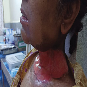

Figure 1. On presentation

Figure 2. Post-procedure day 1

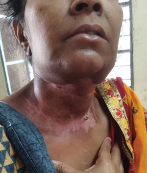

Figure 3. Post-procedure on day 14
Discussion
Acute radiation dermatitis (ARD) may present in the form of erythema, dry and moist desquamation, skin necrosis, ulcers, as well as bleeding. The Radiation Therapy Oncology Group (RTOG) and the European Organization for Research and Treatment of Cancer (EORTC) have developed a standardized grading system to evaluate acute radiation-induced skin toxicity (Table 1) [5].
A measurement tool has also been created that helps evaluate ARD using both patient symptoms and healthcare professionals’ assessment scales. The tool is called the Radiation-Induced Skin Reaction Assessment Scale (RISRAS) [6]. Another important classification of ARD more frequently used in clinical trials than RTOG/EORTC scale is Common Terminology Criteria for Adverse Events (CTCAE) version 5.0 (Table 1).
Table 1. The classification of acute radiation dermatitis
|
Grade |
RTOG/EORTC |
CTCAE 5.0 |
|---|---|---|
|
0 |
No change over baseline |
N/A |
|
1 |
Follicular, faint, or dull erythema; epilation; dry desquamation; decreased sweating |
Faint erythema or dry desquamation |
|
2a |
Tender or bright erythema ± dry |
Moderate-to-brisk erythema; patchy moist desquamation, mostly confined to skin folds and creases; moderate edema |
|
2b |
Patchy moist desquamation; moderate edema |
|
|
3 |
Confluent moist desquamation other than skin folds; pitting edema |
Moist desquamation in areas other than skin folds and creases; bleeding induced by minor trauma or abrasion |
|
4 |
Ulceration, haemorrhage, necrosis |
Life-threatening consequences; skin necrosis or ulceration of full-thickness dermis; spontaneous bleeding from the involved site; skin graft indicated |
|
5 |
N/A |
Death |
Several dressings were investigated in both ARD prophylaxis and treatment. The main aim of their application is the reduction of ARD severity or severity of related symptoms (such as pain) and subsequent improvement of treatment tolerance [7]. Clinically, immediate coverage of the open burn wound is necessary to ensure a satisfactory outcome. This is the most important factor determining the recovery of the patient.
The amniotic membranes, being thin film dressings, are easy to use clinically. The amniotic membrane consists of extracellular matrix (ECM), biologically energetic cells, collagen and regenerative molecules. ECM provides framework and contains large amounts of proteins, such as laminins, fibronectin, and proteoglycans, which along with collagen provide structural strength to the membrane. The biologically energetic cells include stem cells, whose function it is to regenerate the latest cellular substances to the coating of the membrane. Fibroblasts help to strengthen the tissue, and the epithelial cells aid in the healing process mediated by the receptors present on the cellular surface. Regenerative molecules are necessary for healing and growth. It includes fibroblast growth factors, platelet-derived growth factors, and metalloproteinase. In a study by Lobo Gajiwala and Sharma [8], the cryopreserved human amniotic membrane has been successfully used in the management of moist desquamations. It can conform to the area of moist desquamation and provide an effective barrier to trauma and microbial penetration. It also helps in retaining a physiologically moist microenvironment which promotes healing. Amniotic membrane promotes epithelialization and hence decreases the need for multiple dressings [9]. It is contraindicated in the presence of infection. They are also easy to apply onto the burn surface due to their good conformability. It adhered firmly to the wound. They become dry membranes covering the wound and protect the wound from contamination and infection. Amniotic membranes are not rejected by the host immune system.
Advantages of amnion membranes are:
• easy to procure and process (ease of storage and long shelf-life);
• cheap and cost effective (reduces length of hospital stay);
• reduces contamination and infection rate;
• reduces need for intensive nursing care;
• potential for use in general surgical or burn ward;
• potential for use in peripheral or district hospitals;
• availability of good quality amnion from hospitals with a high delivery rate.
Albeit its efficacy in successful wound management, its use is limited due to poor availability.
Conclusively, proper patient selection and usage of amniotic membranes with long shelf-life, has an unequivocal potential in the treatment of acute post radiation burn wounds. Its clinical usage as a burn wound dressing has been proven beyond doubt. It is of great significance in the management of burns in children and teenagers, especially for the alleviation of pain. It has an added advantage of single application to the wound without the need for re-dressing the burn wound. The average healing time is about 6 weeks. Disadvantageously, the smell associated with dressing and the acceptability of human placenta as a dressing are limiting its use. Analytically, given the advantages and effective healing, the associated negative factors have a very poor leverage. Thus, amniotic membranes can serve as a potential dressing for management of acute post radiation burns.
Author contributions
Lohit Tejasvi conceived the concept, accessed clinical data related to the patients, and obtained the data.
Ayjaz Hussain drafted the initial manuscript, handled data analysis, literature review, and editing of the manuscript.
All the authors read and approved the final manuscript.
References
1. Rosenthal A, Israilevich R, Moy R. Management of acute radiation dermatitis: A review of the literature and proposal for treatment algorithm. J Am Acad Dermatol 2019; 81(2): 558‒567.
2. Bray FN, Simmons BJ, Wolfson AH, Nouri K. Acute and chronic cutaneous reactions to ionizing radiation therapy. Dermatol Ther 2016; 6(2): 185–206.
3. Spałek M. Chronic radiation-induced dermatitis: Challenges and solutions. Clin Cosmet Investig Dermatol 2016; 9: 473–482.
4. Saini S, Pai V, Shukla P, Ranglani H. A study of clinical pattern of acute radiation dermatitis among patients attending dermatology outpatient department at tertiary center in Western India. Clin Dermatol Rev 2018; 2(1): 8‒12.
5. Cox JD, Stetz J, Pajak TF. Toxicity criteria of the Radiation Therapy Oncology Group (RTOG) and the European Organization for Research and Treatment of Cancer (EORTC). Int J Radiat Oncol Biol Phys 1995; 31(5): 1341–1346.
6. Noble-Adams R. Radiation-induced skin reactions. 2: Development of a measurement tool. Br J Nurs 1999; 8(18): 1208–1211.
7. Zasadziński K, Spałek MJ, Rutkowski P. Modern dressings in prevention and therapy of acute and chronic radiation dermatitis ‒ a literature review. Pharmaceutics 2022; 14(6): 1204.
8. Lobo Gajiwala A, Sharma V. Use of irradiated amnion as a biological dressing in the treatment of radiation induced ulcers. Cell Tissue Bank 2003; 4(2‒4): 147‒150.
9. Malhotra C, Jain AK. Human amniotic membrane transplantation: Different modalities of its use in ophthalmology. World J Transplant 2014; 4(2): 111‒121.