Lietuvos chirurgija ISSN 1392–0995 eISSN 1648–9942
2026, vol. 25(1), pp. 70–73 DOI: https://doi.org/10.15388/LietChirur.2026.25(1).8
Congenital Anterior Urethrocutaneous Fistula: A Rare Case Report
Neel Aggerwal
Post Graduate Institute of Child Health, Department of Pediatric Surgery, Noida, India
E-mail: aggerwal.pgich@gmail.com
Chandra Vibhash
Post Graduate Institute of Child Health, Department of Pediatric Surgery, Noida, India
E-mail: chandravibhash@yahoo.com
Pragya Mishra
Post Graduate Institute of Child Health, Department of Pediatric Surgery, Noida, India
E-mail: tinytots2417@gmail.com
Umesh B. Singh
Post Graduate Institute of Child Health, Department of Pediatric Surgery, Noida, India
E-mail: singhub.2008@gmail.com
Abstract. Congenital anterior urethrocutaneous fistula (CAUF) is an extremely rare penile anomaly which presents despite a normal urethral meatus and intact prepuce. It may present in isolation or with associated anomalies such as hypospadias or chordee. We report the case of a 14-months-old boy with a congenital anterior urethrocutaneous fistula on the ventral penile shaft. A single-stage urethrocutaneous fistula closure with a dartos flap along with torsion correction was performed. The postoperative course was uneventful, and the child remains well on follow-up.
Keywords: congenital, urethrocutaneous fistula, penile anomaly.
Received: 2025-08-17. Accepted: 2025-09-25.
Copyright © 2026 Neel Aggerwal, Chandra Vibhash, Pragya Mishra, Umesh B. Singh. Published by Vilnius University Press. This is an Open Access article distributed under the terms of the Creative Commons Attribution Licence, which permits unrestricted use, distribution, and reproduction in any medium, provided the original author and source are credited.
Introduction
Congenital anterior urethrocutaneous fistula (CAUF) is an uncommon penile anomaly characterized by an abnormal urethral opening on the ventral shaft, which has a normal meatus and intact prepuce. It may present with or without hypospadias and chordee. It may also occur as an isolated defect or in association with other genitourinary anomalies. The precise aetiology is uncertain but likely stems from a focal developmental defect in the urethral plate, preventing complete fusion of the urethral folds [1].
Case report
A 14-month-old male child presented with urine leakage from the underside of the penis for two months. Since birth, the child voided normally from the urethral meatus, although a hypopigmented patch was noted on the ventral penile shaft. Two months prior to presentation, the parents observed urine dribbling from a pinpoint opening within the discoloured patch, though child kept passing urine in good stream from glanular meatus. There was no history of trauma, surgery, or similar familial anomalies.
Systemic examination was normal. On local examination bilateral testis were adequate for age and well placed in scrotum. Stretched penile length was satisfactory for age, with a normal well-developed look and intact prepuce. Ventral shaft of penis had a 1×1 cm hypopigmented patch just proximal to the coronal sulcus, with a 5×5 mm fistulous opening. This patch was seen adhered to underlying tissue and the penile skin was not free around it. The patch and urethra-cutaneous fistula lied on right side of the mid penile raphe, with 30-degree torsion seen to right (Figure 1). The glans was intact with a normal urethral meatus and prepuce. There was no chordee and the anus was normal. No other anomalies were appreciated on detailed examination.
The patient underwent surgical repair under general anaesthesia. When calibrated with 8 Fr infant feeding tube, it passed easily from glanular meatus to the bladder. Thinned out tissues and urethra were seen at patch area with almost visible infant feeding tube, but glans and coronal area distal to UCF was seen intact with no tissue dehiscence (Figure 1).
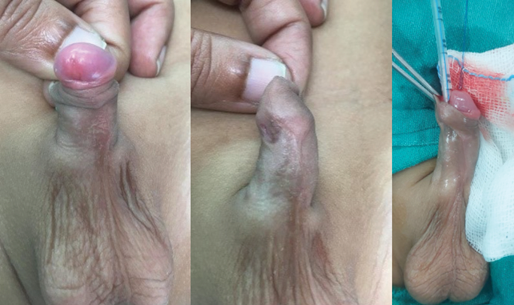
Figure 1. Pre-operative images
The goal of surgery, henceforth, was to release the torsion and close the UCF with preserving already well-formed tissues. The patch and UCF were dissected out all around from underlying tissue like usual UC fistula closure and the skin of the penis was degloved completely till the torsion was released. After the fistula was closed over itself, a dartos flap was interposed to reinforce the repair. Skin was covered after complete repair with Byar’s flap. The glanular urethra and meatus were left intact (Figure 2). Recovery was uneventful, and the patient is doing well on follow-up.

Figure 2. Post-operative images
Discussion
Primary anterior CAUF is extremely rare, with fewer than 70 cases described in the literature till date. A systematic review by Lin et al. (1962–2017) identified 63 cases, with the subcoronal location being most common, followed by mid-penile and penoscrotal sites [2].
Two anatomical variants of CAUF have been described: one with a hypoplastic distal urethra resembling proximal hypospadias, and the other ‒ seen in our patient ‒ with a normal urethra proximal and distal to the fistula [1]. The pathogenesis remains speculative; Campbell suggested these represent embryological urethral blowouts due to distal obstruction [3].
Diagnosis relies on clinical history and examination. A history of prior trauma or circumcision must be ruled out. As sometimes urethrocutaneous fistula is seen post circumcision due to damage to the urethra in the process. But in our case the prepuce was intact and there was no history of any surgery performed let alone circumcision. Imaging such as ultrasound and voiding cystourethrogram (VCUG) may help assess for associated anomalies or confirm dual urinary streams [4].
Multiple surgical techniques have been used to correct CAUF, including local skin flaps, Thiersch-Duplay or Denis Browne urethroplasty, and buccal mucosal grafts. Tubularized incised plate urethroplasty with dartos flap cover offers excellent outcomes, especially when the distal urethra is intact. The choice of technique should be individualized based on fistula location, size, and associated anomalies [5].
Conclusion
Congenital anterior urethrocutaneous fistula is a rare but correctable anomaly. A tailored surgical approach based on anatomical findings yields favourable outcomes. Preoperative evaluation for associated anomalies is essential, and hypospadias repair principles should guide surgical planning.
Author contributions
Neel Aggerwal ‒ conceptualization, investigations, writing original draft.
Chandra Vibhash ‒ conceptualization, writing original draft.
Pragya Mishra ‒ investigations, writing review and editing.
Umesh B. Singh ‒ visualization, writing review and editing.
References
1. Bhatnagar A, Upadhyaya V, Kumar B. Congenital urethrocutaneous fistula: Case report with review of literature. Indian J Plast Surg 2012; 45(3): 563–565.
2. Lin YW, Yang SS, Hsieh CH, Chang SJ. Congenital anterior urethrocutaneous fistula: A systematic review of 63 cases. Pediatr Surg Int 2017; 33(4): 435–441.
3. Campbell MF, Harrison JH. Campbell’s urology. 5th ed. Philadelphia: WB Saunders, 1986, p. 1873–1880.
4. Zhang B, Zhong H, Geng H, Zhang B. Clinical features and outcomes of congenital anterior urethrocutaneous fistula: A case series study. Urol Int 2025; 109(5): 528‒532.
5. Alghamlas A, Auber F, Chaussy Y. Congenital urethral fistula: A case report and literature review. Case Rep Urol 2020; 2020: 8862806.