Lietuvos chirurgija ISSN 1392–0995 eISSN 1648–9942
2026, vol. 25(2), pp. 176–181 DOI: https://doi.org/10.15388/LietChirur.2026.25(2).8
Diagnostic Challenges in Differentiating Multiple Primary Lung Cancer from Intrapulmonary Metastasis: A Clinical Case Report
Rimantė Šalkauskaitė
Vilniaus universitetas, Medicinos fakultetas, Klinikinės medicinos institutas,
Krūtinės ligų, imunologijos ir alergologijos klinika, Vilnius, Lietuva
Vilnius University, Faculty of Medicine, Institute of Clinical Medicine,
Clinic of Chest Diseases, Immunology, and Allergology, Vilnius, Lithuania
El. paštas rimante.salkauskaite@mf.stud.vu.lt
https://orcid.org/0009-0001-9244-9432
https://ror.org/03nadee84
Agnė Šimbelytė
Vilniaus universitetas, Medicinos fakultetas, Biomedicinos mokslų institutas,
Patologijos, teismo medicinos katedra, Vilnius, Lietuva
Vilnius University, Faculty of Medicine, Institute of Biomedical Sciences,
Department of Pathology and Forensic Medicine, Vilnius, Lithuania
El. paštas agne.simbelyte@vpc.lt
https://ror.org/03nadee84
Vytautas Jovaišas
Vilniaus universitetas, Medicinos fakultetas, Klinikinės medicinos institutas,
Krūtinės ligų, imunologijos ir alergologijos klinika, Vilnius, Lietuva
Vilnius University, Faculty of Medicine, Institute of Clinical Medicine,
Clinic of Chest Diseases, Immunology, and Allergology, Vilnius, Lithuania
Vilniaus universiteto ligoninė Santaros klinikos, Širdies ir krūtinės chirurgijos centras,
Krūtinės chirurgijos skyrius, Vilnius, Lietuva
Vilnius University Hospital Santaros Klinikos, Center of Cardiothoracic Surgery,
Department of Thoracic Surgery, Vilnius, Lithuania
El. paštas vytautas.jovaisas@santa.lt
https://ror.org/03nadee84
Vytenis Bertašius
Vilniaus universitetas, Medicinos fakultetas, Klinikinės medicinos institutas,
Krūtinės ligų, imunologijos ir alergologijos klinika, Vilnius, Lietuva
Vilnius University, Faculty of Medicine, Institute of Clinical Medicine,
Clinic of Chest Diseases, Immunology, and Allergology, Vilnius, Lithuania
Vilniaus universiteto ligoninė Santaros klinikos, Širdies ir krūtinės chirurgijos centras,
Krūtinės chirurgijos skyrius, Vilnius, Lietuva
Vilnius University Hospital Santaros Klinikos, Center of Cardiothoracic Surgery,
Department of Thoracic Surgery, Vilnius, Lithuania
El. paštas vytenis.bertasius@santa.lt
https://ror.org/03nadee84
Žymantas Jagelavičius
Vilniaus universitetas, Medicinos fakultetas, Klinikinės medicinos institutas,
Krūtinės ligų, imunologijos ir alergologijos klinika, Vilnius, Lietuva
Vilnius University, Faculty of Medicine, Institute of Clinical Medicine,
Clinic of Chest Diseases, Immunology, and Allergology, Vilnius, Lithuania
Vilniaus universiteto ligoninė Santaros klinikos, Širdies ir krūtinės chirurgijos centras,
Krūtinės chirurgijos skyrius, Vilnius, Lietuva
Vilnius University Hospital Santaros Klinikos, Center of Cardiothoracic Surgery,
Department of Thoracic Surgery, Vilnius, Lithuania
El. paštas zymantas.jagelavicius@mf.vu.lt
https://orcid.org/0000-0002-6344-8596
https://ror.org/03nadee84
Abstract. Background. Multiple primary lung cancers increasingly emerge in clinical practice due to improved imaging, prolonged patient survival, and growing recognition of this condition. Distinguishing multiple primary lung cancers from intrapulmonary metastases remains challenging and directly influences staging and treatment strategy. Case presentation. A 72-year-old long-term smoker was incidentally found to have a left-lung squamous cell carcinoma and additional right-lung lesions. Initial radiological and histological evaluations suggested separate primary tumors, leading to left upper lobectomy and wedge resections of two right-lung nodules. Histopathology revealed pleomorphic carcinoma in the left lung, squamous cell carcinoma in the right S9 segment, and adenocarcinoma in the S2 segment. Later reassessment classified the squamous carcinoma as a metastasis. The patient received adjuvant carboplatin–paclitaxel chemotherapy and remained recurrence-free after 4.5 years of follow-up. Conclusion. This case emphasizes the complexity of multiple primary lung cancer diagnoses and the need for a multidisciplinary approach integrating imaging, histopathology, and molecular methods. Accurate differentiation is essential for optimal management.
Keywords: multiple primary lung cancer, intrapulmonary metastasis, differential diagnosis, molecular profiling, lung cancer treatment.
Diagnostiniai iššūkiai diferencijuojant daugybinius pirminius plaučių navikus ir plaučių metastazes: klinikinis atvejis
Santrauka. Įvadas. Klinikinėje praktikoje vis dažniau susiduriama su daugybiniais pirminiais plaučių navikais, nes gerėja vaizdinių tyrimų galimybės, ilgėja pacientų išgyvenamumas ir apie šią ligą žinoma daugiau. Daugybinių pirminių plaučių navikų ir intrapulmoninių metastazių diferenciacija, turinti tiesioginę įtaką nustatant ligos stadiją ir parenkant gydymo strategiją, išlieka iššūkiu. Klinikinis atvejis. 72 m. pacientui, daug metų rūkančiam, atsitiktinai nustatytas darinys kairiajame plautyje ir du papildomi dariniai dešiniajame plautyje. Pirminiai radiologiniai ir histologiniai tyrimai parodė atskirus pirminius nesmulkiųjų ląstelių plaučių navikus, todėl atlikta kairioji viršutinė lobektomija ir dviejų dešiniojo plaučio židinių pleištinė rezekcija. Histologinis tyrimas atskleidė pleomorfinę karcinomą kairiajame plautyje, plokščialąstelinę karcinomą dešiniojo plaučio S9 segmente ir adenokarcinomą S2 segmente. Vėliau, tyrimo rezultatus pakartotinai įvertinus tarpdisciplininei komandai, dešiniojo plaučio plokščialąstelinės karcinomos židinys pripažintas kairiojo plaučio naviko metastaze. Pacientui skirta adjuvantinė chemoterapija. Vyras stebėtas 4,5 metų. Recidyvo ar ligos atsinaujinimo požymių nenustatyta. Išvada. Klinikinis atvejis pabrėžia daugybinių pirminių plaučių navikų diagnostikos sudėtingumą ir daugiadalykio vertinimo būtinybę, integruojant vaizdinius, histopatologinius ir molekulinius tyrimus. Tiksli diferenciacija būtina siekiant tinkamiausio gydymo.
Reikšminiai žodžiai: daugybiniai pirminiai plaučių navikai, intrapulmoninė metastazė, diferencinė diagnostika, plaučių vėžio gydymas.
Received: 2026-01-19. Accepted: 2026-02-18.
Copyright © 2026 Rimantė Šalkauskaitė, Agnė Šimbelytė, Vytautas Jovaišas, Vytenis Bertašius, Žymantas Jagelavičius. Published by Vilnius University Press. This is an Open Access article distributed under the terms of the Creative Commons Attribution Licence, which permits unrestricted use, distribution, and reproduction in any medium, provided the original author and source are credited.
Background
Multiple primary lung cancers (MPLC) are defined as two or more synchronous or metachronous primary malignant tumors occurring in the lungs [1]. Primary multiple lung cancers are classified as synchronous (sMPLC) when at least two primary tumors are present simultaneously within the same lung or the contralateral lung within a six-month interval. In contrast, metachronous MPLC (mMPLC) refers to cases in which the second primary tumor is diagnosed more than six months after the first [2].
The reported incidence of sMPLC in various clinical series ranges from 0.2 to 8% (3.5–14% in autopsy studies). The number of diagnosed MPLC cases has been increasing in recent years due to improved early detection through computed tomography and positron emission tomography, longer survival of lung cancer patients, and growing recognition of this clinical phenomenon [3].
According to the Martini and Melamed criteria, tumors with different histological subtypes can be classified as separate tumors; however, more than 50% of lesions may share identical histology [3]. Therefore, comprehensive diagnostic evaluation using diagnostic imaging tools is crucial to differentiate metastases from primary tumors, as this distinction guides therapeutic decision-making and has prognostic implications [4].
To demonstrate the difficulty of distinguishing separate primary tumors from metastatic spread, we report a case involving multifocal pulmonary lesions.
Case report
A 72-year-old man with a 50-year smoking history was incidentally found to have a lesion in the left lung (Figure 1). Transbronchial biopsy revealed a poorly differentiated (G3) squamous cell carcinoma with low PD-L1 expression (1‒49%). Whole-body PET imaging demonstrated additional metabolically active lesions in the right lung (segments S9 and S2) (Figure 2). Biopsy of the larger S9 lesion confirmed a poorly differentiated G3 squamous cell carcinoma with low PD-L1 expression (1‒49%). Transbronchial lymph node biopsy under endobronchial ultrasound guidance was performed to accurately stage the disease and differentiate between two independent primary tumors and metastasis. It showed no evidence of nodal involvement.
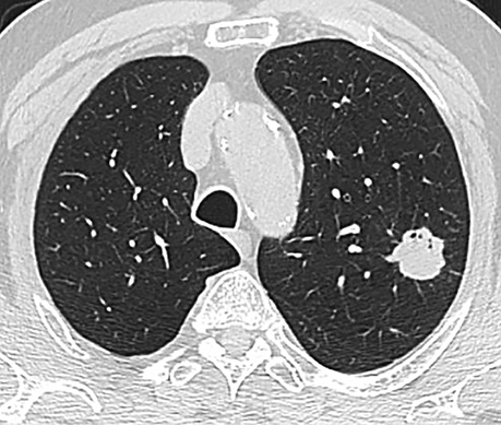
Figure 1. Chest CT scan – primary lesion in the upper lobe of the left lung
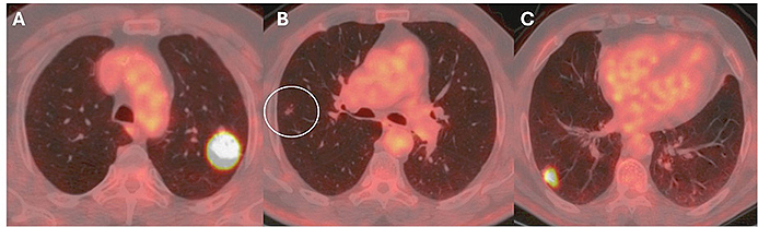
Figure 2. Chest PET‒CT scan showing metabolically active lesions in the left lung (A), and in the right lung (B, segment S9; C, segment S2)
The tumors were initially considered as separate primary lesions, which indicated a choice of radical treatment. The patient underwent left upper lobectomy with lymphadenectomy. Histological examination revealed a poorly differentiated G3 pleomorphic carcinoma (squamous and spindle-cell components), staged as pT2aN0M0, IB. After one month, a wedge resection of the right lung followed. Two lesions in segments S9 and S3 were removed. Histological examination of the S9 lesion revealed a poorly differentiated G3 squamous cell carcinoma with parenchymal invasion, staged as pT1cN0M0, IA3. The S3 lesion was identified as a moderately differentiated G2 adenocarcinoma (pT1aN0M0, IA1) (Figure 3).
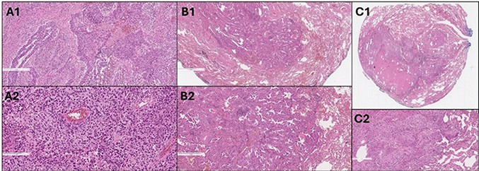
Figure 3. Histological examination. Pleomorphic carcinoma: squamous cell carcinoma part (A1), spindle cell carcinoma part (A2). Adenocarcinoma (B1, B2). Squamous cell carcinoma (C1, C2).
Left lung tumor and one right lung lesion shared identical histology; therefore, in a multidisciplinary discussion, it was reclassified as synchronous disease consisting of a right lung adenocarcinoma (pT1a, stage IA1) and a left lung pleomorphic carcinoma staged as T2aN0M1a (stage IVA) due to metastasis to the right lung. Accordingly, adjuvant chemotherapy for squamous cell carcinoma was initiated. The patient received four cycles of intravenous carboplatin and paclitaxel.
During 4.5 years of follow-up, the disease remained radiologically stable without signs of recurrence.
Discussion
This clinical case illustrates the complexity and clinical significance of differentiating primary tumors from metastases. Accurate diagnosis determines staging, therapeutic strategies, and prognosis.
The earliest diagnostic framework, developed by Martini and Melamed, is based on clinical, radiological, and histopathological criteria that distinguish MPLC from intrapulmonary metastases (IPM). According to these guidelines, differences in tumor histology, distinct anatomical location, and absence of distant metastases are key features supporting MPLC [5]. Although histological assessment is essential, multiple studies highlight its moderate sensitivity and specificity in differentiating MPLC from IPM [6], particularly when the tumors share similar histopathologic features [7].
In the present case, the absence of lymph node involvement initially supported classification of the right-lung lesion as a separate primary tumor despite similar histology. Given the tumor sizes and the patient’s operative fitness, surgical management was selected.
Surgical resection remains the main treatment for operable MPLC patients [1, 8]. Lobectomy is typically preferred; however, limited resections are considered safe, particularly when multiple lung regions are affected or postoperative pulmonary reserve is limited. Increasing emphasis is placed on parenchyma-sparing techniques such as segmentectomy or wedge resection, especially for patients with expected long-term survival or an elevated risk of additional primary tumors (e.g., smokers, patients with prior MPLC) [9].
Nevertheless, uncertainty remains regarding whether the right-lung lesion represented a metastasis or an independent primary tumor with identical histology. In such cases, molecular profiling such as next-generation sequencing could improve differentiation by assessing genetic signatures. Tumors with distinct molecular profiles can be classified as separate primaries; in contrast, highly concordant profiles suggest metastasis. However, the limitations of molecular testing include tumor heterogeneity, which may complicate interpretation, especially when only small biopsy samples are available [7].
Conclusions
The optimal strategy to distinguish MPLC from IPM requires a multidisciplinary approach integrating clinical data, radiological findings, histological evaluation, and molecular diagnostics. Histology provides morphological features, imaging adds anatomical context, and molecular methods reveal genomic characteristics critical for precise classification. Although accurate differentiation is crucial for determining treatment and prognosis, definitive classification of all multifocal lung tumors remains challenging.
Author contribution
Rimantė Šalkauskaitė ‒ conceptualization, data collection, writing original draft, visualization, and literature review.
Agnė Šimbelytė – histological images and histological comments, general review.
Vytautas Jovaišas – general manuscript review and editing.
Vytenis Bertašius – general manuscript review and editing, literature review.
Žymantas Jagelavičius ‒ conceptualization, supervision, editing images, manuscript review, and editing.
Acknowledgements. Not applicable.
Conflict of interest. No conflicts of interest.
Funding. This work was not supported by any funding source.
Ethical approval. Not required.
Consent. The patient gave written permission for the publication of this case report and any accompanying images.
References
1. Chen C, Huang X, Peng M, Liu W, Yu F, Wang X. Multiple primary lung cancer: A rising challenge. J Thorac Dis 2019; 11(Suppl 4): S523‒S536.
2. Huimin Z, Xueting W, Qi Q, Lingxin F, Xue Y, Zhuang Y, Jing W. Multiple primary lung cancers with ALK rearrangement: A case report and literature review. Front Oncol 2022; 12: 897451.
3. Loukeri AA, Kampolis CF, Ntokou A, Tsoukalas G, Syrigos K. Metachronous and synchronous primary lung cancers: Diagnostic aspects, surgical treatment, and prognosis. Clin Lung Cancer 2015; 16(1): 15–23.
4. Stella F, Luciano G, Dell’Amore A, Greco D, Ammari C, Giunta D, Bini A. Pulmonary metastases from NSCLC and MPLC (Multiple Primary Lung Cancers): Management and outcome in a single centre experience. Heart Lung Circ 2016; 25(2): 191–195.
5. Bai G, Li Y, Ji Y, Peng Y, Yang Z, Zhao L. Treatments and whole exon sequencing of a case with multiple primary lung cancer. J Cardiothorac Surg 2023; 18(1): 58.
6. Tian H, Bai G, Yang Z, Chen P, Xu J, Liu T, Fan T, Wang B, Xiao C, Li C, Gao S, He J. Multiple primary lung cancer: Updates of clinical management and genomic features. Front Oncol 2023; 13: 1034752.
7. Olteanu GE, Kern I, Kalson L, Brcic L. From morphology to molecules: Advances in the distinction of multiple primary lung cancers from intrapulmonary metastases in non-small cell lung cancer. Adv Anat Pathol 2024; 31(5): 318‒323.
8. Chiang CL, Tsai PC, Yeh YC, Wu YH, Hsu HS, Chen YM. Recent advances in the diagnosis and management of multiple primary lung cancer. Cancers 2022; 14(1): 242.
9. Zhao L, Liu C, Xie G, Wu F, Hu C. Multiple primary lung cancers: A new challenge in the era of precision medicine. Cancer Manag Res 2020; 12: 10361–10374.