Lietuvos chirurgija ISSN 1392–0995 eISSN 1648–9942
2026, vol. 25(3), pp. 208–218 DOI: https://doi.org/10.15388/LietChirur.2026.25(3).1
Priapism: A Comprehensive Review of Pathophysiology, Evidence-Based Management, and Future Directions
Muhammad Munir Memon
Qassim University, College of Medicine, Department of Surgery, Qassim, Saudi Arabia
Qassim University, Medical City, Department of General Surgery, Qassim, Saudi Arabia
E-mail: m.ghafar@qu.edu.sa
https://orcid.org/0000-0003-2501-3126
Emad Abdulrhamn S. AlWashmi
Qassim University, College of Medicine, Department of Surgery, Qassim, Saudi Arabia
Qassim University, Medical City, Department of General Surgery, Qassim, Saudi Arabia
Qassim University, Medical City, Department of Urology and Assisted Reproductive Technology, Buraydah, Saudi Arabia
E-mail: E.Alwashmi@qu.edu.sa
Abstract. Priapism, defined as a persistent penile erection lasting beyond four hours unrelated to sexual stimulation, is a urological emergency with significant implications for long-term sexual function. Its most common form, ischemic priapism (IP), represents a compartment syndrome of the penis, necessitating immediate intervention to prevent corporal fibrosis and permanent erectile dysfunction. Despite the potentially devastating complications of this condition, management of recurrent priapism episodes historically has commonly involved reactive treatments rather than preventative strategies. This comprehensive narrative review provides a critical analysis of the contemporary understanding of priapism. We delve into the molecular and hemodynamic mechanisms underlying the refined pathophysiological classification: ischemic (low-flow), non-ischemic (high-flow), and stuttering (recurrent) priapism. Recently, increasing elucidation of the complex molecular mechanisms underlying this disorder, principally involving dysregulation of nitric oxide signaling, has allowed for greater insights and exploration into potential therapeutic targets. In this review, we discuss the multiple molecular regulatory pathways implicated in the pathophysiology of priapism. We also identify the roles and mechanisms of molecular effectors in providing the basis for potential future therapies. The review underscores the critical importance of a rapid, systematic diagnostic approach utilizing history, examination, corporal blood gas analysis, and color Doppler ultrasonography. Management is meticulously detailed, emphasizing the stepwise, escalating protocol for IP, from aspiration and sympathomimetic irrigation to surgical shunting, and the conservative or minimally invasive strategies for non-ischemic priapism. A significant focus is placed on the paradigm shift in managing stuttering priapism, where targeted therapies, including the paradoxical use of phosphodiesterase type 5 (PDE5) inhibitors, are revolutionizing prevention. We also explore special populations, long-term sequelae, and future directions, including novel molecular targets, advanced imaging, and regenerative medicine. The objective is to equip clinicians with a sophisticated, evidence-based framework to optimize outcomes in this challenging clinical scenario.
Keywords: priapism, ischemic priapism, erectile dysfunction, sickle cell disease, pathophysiology, corporal aspiration, surgical shunt, PDE5 inhibitors.
Received: 2026-03-12. Accepted: 2026-04-16.
Copyright © 2026 Muhammad Munir Memon, Emad Abdulrhamn S. AlWashmi. Published by Vilnius University Press. This is an Open Access article distributed under the terms of the Creative Commons Attribution Licence, which permits unrestricted use, distribution, and reproduction in any medium, provided the original author and source are credited.
Introduction
Priapism, named after the Greek fertility god Priapus, is a condition that has been documented since antiquity, yet its effective management remains a modern clinical challenge. The formal definition – a full or partial penile erection persisting for more than 4 hours, unrelated to sexual interest or stimulation – belies a complex underlying pathophysiology [1]. With an estimated incidence of 1.5 to 5.34 per 100,000 person-years, it is a relatively rare but true medical emergency [2]. The urgency is driven predominantly by ischemic priapism (IP), which accounts for over 95% of cases and mirrors a compartment syndrome, characterized by rigid erection, pain, and vicious cycle of hypoxia, acidosis, and ultimately, irreversible corporal tissue damage if not promptly reversed [3].
The clinical approach has evolved from a purely mechanical focus on detumescence to a nuanced understanding of molecular signaling pathways that govern penile erection and detumescence. This evolution has been particularly transformative for patients with recurrent (stuttering) priapism, for whom preventive molecular therapies are now a reality. This narrative review aims to provide a critical and comprehensive synthesis of current evidence, from fundamental pathophysiology to advanced management strategies and emerging research, providing a resource for clinicians to navigate this high-stakes condition effectively.
Classification and pathophysiology: A molecular and hemodynamic perspective
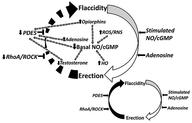
Figure 1. Pathophysiological mechanisms. A diagram comparing IP (showing blocked venous outflow, hypoxic/acidotic blood, and smooth muscle contraction) with NIP (showing an arterio-cavernosal fistula with high-flow, oxygenated blood).
Schematic representation of the molecular pathophysiologic mechanisms of priapism; normal penile erection physiology.
Multidisciplinary management algorithm for pediatric priapism. Reproduced from Song and Moon (2013) under CC BY-NC 3.0 license.
A constellation of molecular factors promotes uncontrolled erection (priapism) by interfering with the normal regulatory control mechanisms involved in the return of the penis back to its flaccid state. Circular arrows represent pathway between penile erection states. Horizontal black arrows represent mediation. Horizontal black T-shapes represent inhibition.
Broken arrows represent both direct and indirect downstream effects of signaling pathways.
Upward black arrows represent upregulation. Downward black arrows represent downregulation.
NO/cGMP – nitric oxide/cyclic guanosine monophosphate, ROS/RNS – reactive oxygen species/reactive nitrogen species, ROCK – rho-associated protein kinase, PDE5 – phosphodiesterase type 5, HO – heme oxygen.
A precise understanding of the pathophysiological classification is not merely academic; it is the direct determinant of management. The classic triad is Ischemic, Non-Ischemic, and Stuttering priapism.
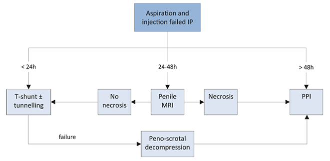
Figure 2. The pathophysiology diagram
Creative Commons Attribution Non-Commercial (CC BY-NC 3.0) Molecular mechanisms of priapism. Reproduced from Song and Moon (2013) under CC BY-NC 3.0 license. Proposed molecular mechanisms of priapism. DOI (https://doi.org/10.4111/kju.2013.54.12.816) serves as the permanent legal identifier for the source. Pathophysiological mechanisms of priapism. Reproduced from Song PH, Moon KH. Priapism: Current updates in clinical management. Korean J Urol 2013; 54(12): 816–823. Licensed under CC BY-NC 3.0.
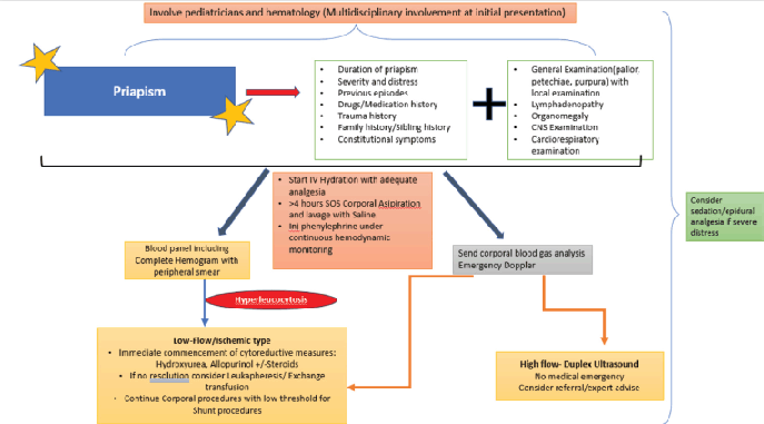
Figure 3. Management algorithm for ischemic priapism
Clinical Management Algorithm for Ischemic and Non-Ischemic Priapism. Adapted from Song and Moon (2013) under CC BY-NC 3.0 license. Management algorithm for failed initial treatment of ischemic priapism. Reproduced from Song and Moon (2013) under CC BY-NC 3.0 license.
Types of priapism
A. Ischemic priapism (low-flow): A compartment syndrome
IP is characterized by a failure of veno-occlusion, leading to stagnant blood within the corpora cavernosa. The resulting environment is hypoxic (pO2 < 30 mmHg), acidotic (pH < 7.25), and hypercarbic, triggering a self-perpetuating cycle of smooth muscle dysfunction and cytotoxicity [4, 5].
Molecular pathogenesis. The initial trigger (e.g., intracavernosal vasodilators, sickle cell erythrocytes) leads to excessive and prolonged corporal smooth muscle relaxation. The critical failure lies in the dysregulation of detumescence:
• Excessive vasoconstrictor tone. Hypoxia and acidosis impair the reuptake of norepinephrine from adrenergic nerve endings, leading to prolonged smooth muscle contraction and further veno-occlusion failure [6].
• Ion channel dysfunction. Hypoxia impairs adenosine triphosphate (ATP)-dependent ion pumps (e.g., Ca2+ ATPase). This leads to intracellular calcium ([Ca2+]i) overload, promoting sustained contraction and activating proteolytic enzymes.
• Inflammatory and profibrotic cascade. The hypoxic environment upregulates transforming growth factor-beta (TGF-β), platelet-derived growth factor (PDGF), and other cytokines. This promotes the transformation of corporal smooth muscle cells into a profibrotic phenotype, leading to collagen deposition, apoptosis, and ultimately, corporal fibrosis [7]. The point of no return for significant fibrosis is often considered to be between 24 and 48 hours.
Common etiologies. Hematological dyscrasias (sickle cell disease being the most prominent), intracavernosal pharmacotherapy (e.g., prostaglandin E1, papaverine), antipsychotics (e.g., trazodone, risperidone), and recreational drugs (e.g., cocaine).
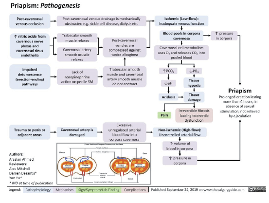
Figure 4. Pathogenesis of ischemic and non-ischemic priapism
Reproduced from Ahmad A. Priapism: Pathogenesis. The Calgary Guide to Understanding Disease (2019). Available at www.thecalgaryguide.com. Licensed under CC BY-NC-SA 4.0.
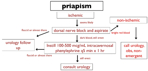
Figure 5. Management algorithm for ischemic priapism
A flowchart starting with “Presentation <4 hrs, Rigid, Painful” leading to Aspiration/Irrigation → Phenylephrine → Distal Shunt → Proximal Shunt, with key decision points.
Initial management algorithm for ischemic and non-ischemic priapism. Schematic detailing the diagnostic and therapeutic steps, including aspiration and intracavernosal phenylephrine. Adapted from the American Urological Association (AUA) Guideline on the Management of Priapism (2003).
B. Non-ischemic priapism (high-flow): Unregulated inflow
NIP is typically post-traumatic, resulting from an arterio-cavernosal fistula that bypasses the regulatory helicine arteries. This allows unregulated, high-flow arterial blood directly into the lacunar spaces [8]. Crucially, the blood remains well-oxygenated (pO2 > 90 mmHg, pH ~ 7.4), meaning there is no tissue ischemia or immediate risk of fibrosis. The erection is typically painless, partial, and compressible.
High-flow (non-ischemic) priapism
• Due to unregulated arterial blood flow, presenting with a semi-rigid, painless erection.
• Usually due to trauma and subsequent fistula development and usually self-limiting.
• Blood gas analysis shows similar results to arterial blood.
Recurrent (or stuttering) priapism
• Most commonly seen in sickle cell disease.
• Usually high flow, but may change to low flow with anoxia.
C. Stuttering (intermittent) priapism: A dysregulation of signaling
This recurrent form of IP is characterized by painful episodes that resolve spontaneously but cause significant distress and carry a cumulative risk of fibrosis. It is highly prevalent in SCD, with a lifetime prevalence of up to 35–45% [9]. The pathophysiology involves a fundamental dysregulation of the nitric oxide (NO)/cyclic guanosine monophosphate (cGMP) pathway. In SCD, chronic hemolysis leads to arginase release, which depletes L-arginine, the substrate for NO synthesis. This creates a state of NO resistance and PDE5 dysregulation, where the normal termination of erection is impaired, leading to uncontrolled, prolonged tumescence [10, 11].
Table 1. Pathophysiological and diagnostic characteristics of priapism subtypes
|
Feature |
Ischemic priapism (IP) |
Non-ischemic priapism (NIP) |
Stuttering priapism |
|---|---|---|---|
|
Pathophysiology |
Veno-occlusive failure, compartment syndrome |
Arterio-cavernosal fistula (usually traumatic) |
Dysregulated NO/cGMP/PDE5 signaling |
|
Blood flow |
Low or absent |
High |
Intermittent IP episodes |
|
Blood gas (CBG) |
Hypoxic (pO2 < 30 mmHg), acidotic (pH < 7.25) |
Normoxic (pO2 > 90 mmHg), normal pH (~7.4) |
Hypoxic during |
|
Clinical presentation |
Rigid, painful, tender |
Soft, painless, non-rigid |
Recurrent, painful, |
|
Color doppler US |
Absent or minimal diastolic flow |
Focal “blush”, high diastolic flow, visible fistula |
Varies; during episode, resembles IP |
|
Treatment urgency |
Urological emergency |
Not an emergency |
Preventive focus |
Diagnostic evaluation: A systematic approach
A rapid, targeted evaluation is paramount.
• History. Elicit duration, pain, history of trauma, SCD, medication review (prescription and recreational), and previous episodes.
• Physical examination. Focus on penile rigidity (rigid corpora cavernosa vs. soft) and tenderness. A rigid shaft with a soft glans/spongiosum is classic for IP; a universally soft or partially rigid penis suggests NIP.
• Corporal blood gas (CBG) aspiration. The definitive bedside test. Aspiration of dark, deoxygenated blood confirms IP. Bright red, well-oxygenated blood is diagnostic of NIP [12].
• Color Doppler ultrasonography. The imaging modality of choice. It objectively confirms hemodynamics: absent flow in IP versus a localized high-flow “blush” or pseudoaneurysm in NIP, often allowing for fistula localization [13].
Evidence-based management strategies
Management is strictly subtype-dependent.
A. Management of ischemic priapism: The escalating ladder
Time is tissue. The goal is to relieve ischemia, reverse acidosis, and restore venous outflow.
First-line (Step 1): Therapeutic aspiration and irrigation
• Aspiration. Using a 19–21-gauge butterfly needle, 20–30 mL of blood is aspirated from each corpus cavernosum. This reduces intracavernosal pressure and removes anoxic blood.
• Irrigation. Gentle irrigation with 0.9% normal saline helps flush out acidic metabolites, vasoactive mediators, and further decompresses the corpora.
Second-line (Step 2): Intracavernosal sympathomimetics
If aspiration/irrigation fails, the next step is intracavernosal injection of an alpha-adrenergic agonist.
• Agent of choice: Phenylephrine (a selective α1-agonist) due to its minimal beta-adrenergic cardiac effects. It is diluted to 100–500 mcg/mL.
• Dosing. 200–500 mcg injections every 3–5 min for up to one hour [14].
• Monitoring. Continuous blood pressure and heart rate monitoring is mandatory to manage potential hypertensive crises or reflex bradycardia.
Third-line (Step 3): Surgical shunting
Indicated if medical therapy fails after 1 hour. The choice of shunt is guided by duration and prior interventions.
• Distal shunts. Create a fistula between the corpora cavernosa and the glans penis.
․ Percutaneous (Ebbehøj). Performed with a large-bore biopsy needle.
․ Open (Al-Ghorab). A more definitive procedure involving excision of a 5 mm core of tunica albuginea from the distal corpora. The T-shunt (with or without tunneling) is a modification that may improve success in prolonged cases [15].
• Proximal shunts. Created between the corpora cavernosa and the corpus spongiosum via a perineal approach. This is more invasive and carries a higher risk of ED and urethral injury.
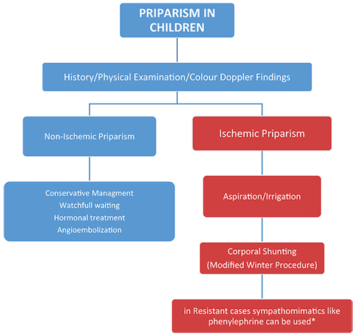
Figure 6. Management algorithm for priapism in children
The flowchart delineates the diagnostic and therapeutic pathways for pediatric patients, distinguishing between non-ischemic (conservative management) and ischemic subtypes (surgical shunting and sympathomimetics). Reproduced from Song PH, Moon KH. Priapism: Current updates in clinical management. Korean J Urol 2013; 54(12): 816–823. Licensed under CC BY-NC 3.0. DOI: 10.4111/kju.2013.54.12.816.
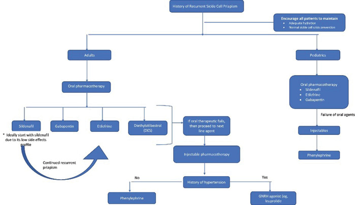
Figure 7. Management algorithm for recurrent sickle cell priapism
Schematic showing the treatment escalation for adults and pediatric patients, including oral and injectable pharmacotherapy. Reproduced from Song PH, Moon KH. Priapism: Current updates in clinical management. Korean J Urol 2013; 54(12): 816–823. Licensed under CC BY-NC 3.0.
Table 2. Stepwise management of ischemic priapism
|
Step |
Intervention |
Procedure |
Key considerations |
|---|---|---|---|
|
1 |
Aspiration and irrigation |
Needle decompression + saline flush |
Diagnostic and therapeutic; may be sufficient for short durations. |
|
2 |
Intracavernosal phenylephrine |
Injections every 3–5 min (max 1 h) |
Continuous cardio-monitoring required. |
|
3a |
Distal surgical shunt |
Percutaneous (Ebbehøj) or open (Al-Ghorab/ |
First-line surgical option. Al-Ghorab/T-shunt is more effective for prolonged priapism. |
|
3b |
Proximal surgical shunt |
Cavernoso-spongiosal anastomosis |
Reserved for distal shunt failure. |
|
Last resort |
Penile prosthesis |
Immediate implantation |
Consider in refractory, prolonged priapism (>48 h) to mitigate complex fibrosis and preserve sexual function. |
B. Management of non-ischemic priapism
Given the absence of ischemia, management is non-urgent. Initial conservative management (observation, ice packs) is justified, as spontaneous resolution occurs in up to 60% of cases [16]. For persistent cases, selective arterial embolization is the gold standard, using absorbable (e.g., gelatin sponge) or non-absorbable (e.g., coils) materials. Absorbable agents have a lower risk of permanent erectile dysfunction. Surgical ligation is reserved for embolization failures.
C. Management of stuttering priapism: A preventive paradigm
The goal is to reduce the frequency and severity of episodes.
• SCD-specific. Optimize hydration, analgesia, and hydroxyurea therapy. Exchange transfusion is reserved for acute episodes, not prophylaxis.
• Pharmacologic prevention:
․ Oral alpha-agonists. Pseudoephedrine has been used anecdotally.
․ Hormonal modulation. Anti-androgens (e.g., finasteride) or GnRH agonists can be effective but have significant side effects (e.g., loss of libido, hot flashes), limiting their use, especially in young men.
․ PDE5 inhibitors (paradoxical use). Daily low-dose sildenafil or tadalafil has demonstrated efficacy in clinical trials for SCD-associated stuttering priapism [17]. The proposed mechanism is the stabilization of the NO/cGMP pathway by reducing oxidative stress and preventing the pathological PDE٥ upregulation that occurs in hypoxic states, thereby promoting normal cyclic erection and detumescence.
Special considerations and long-term sequelae
• Sickle cell disease. A multidisciplinary approach is essential. Management of acute IP follows the standard ladder, but must occur concurrently with SCD crisis management (hydration, oxygenation, analgesia).
• Pediatric priapism. Overwhelmingly associated with SCD. Management requires expertise from pediatric urology and hematology.
• Erectile dysfunction. The most devastating sequela of IP. The risk is directly proportional to the duration of ischemia. All patients must be counseled and offered follow-up. Treatment options range from PDE٥ inhibitors to intracavernosal injections, vacuum devices, and penile implants. In cases of prolonged, refractory priapism, immediate insertion of a penile prosthesis is a viable strategy to create a functional erection and avoid the technical difficulties of implanting into fibrotic corpora at a later date [18].
Future directions and analytical perspectives
The management of priapism is moving from reactive intervention to proactive, molecularly targeted prevention and regeneration.
1. Novel molecular targets
․ Adenosine signaling. Adenosine is a potent vasodilator upregulated in hypoxic states. A2A receptor antagonists are being explored to prevent stuttering priapism [19].
․ RhoA/Rho-kinase (ROCK) pathway. This pathway promotes smooth muscle contraction. ROCK inhibitors may offer a new avenue for treating and preventing priapism.
․ Opiorphins. Endogenous PDE٥ inhibitors; modulating their activity is a theoretical target.
2. Advanced imaging. High-resolution microultrasound and contrast-enhanced ultrasound may provide superior localization of microfistulas in NIP.
3. Regenerative medicine. The ultimate goal for reversing corporal fibrosis. Research into stem cell therapies (e.g., adipose-derived stem cells) aims to repopulate the corpora with healthy smooth muscle and endothelial cells, restoring erectile function [19].
4. Patient-reported outcomes (PROs). Future research must prioritize PROs to fully understand the long-term psychological and quality-of-life impact of priapism and its treatments, guiding more patient-centered care.
Conclusion
Priapism is a complex condition whose successful management hinges on a swift and accurate diagnosis of its underlying subtype. Ischemic priapism demands immediate, aggressive intervention following a stepwise protocol to avert permanent erectile dysfunction. In contrast, non-ischemic priapism permits a deliberate, often minimally invasive approach. The most significant recent advancement lies in the understanding and management of stuttering priapism, where the paradoxical use of PDE5 inhibitors exemplifies a shift towards pathophysiology-driven prevention. As research continues to unravel the intricate molecular dialogues governing penile erection, the future promises even more targeted therapies and regenerative strategies, offering hope for improved quality of life for all patients at risk for this challenging condition.
Author contributions
Muhammad Munir Memon – conducted literature search, data extraction, drafting of the initial manuscript, preparation of tables and figures, and revision of the narrative review.
Emad Abdulrhamn S. AlWashmi – critical review for scientific accuracy, editing of methodology and clinical content, and final approval of the manuscript.
All authors have reviewed and approved the final version of the manuscript.
Ethical approval and informed consent
Since it’s a narrative review article, it does not require ethical approval from IRB.
Conflict of interest
The authors declared no potential conflicts of interest with respect to the research, authorship, and/or publication of this article.
References
1. Montague DK, Jarow J, Broderick GA, Dmochowski RR, Heaton JP, Lue TF, Nehra A, Sharlip ID; Members of the Erectile Dysfunction Guideline Update Panel; Americal Urological Association. American Urological Association guideline on the management of priapism. J Urol 2003; 170(4 Pt 1): 1318–1324. DOI: 10.1097/01.ju.0000087608.07371.
2. Hudnall M, Reed-Maldonado AB, Lue TF. Advances in the understanding of priapism. Transl Androl Urol 2017; 6(2): 199–206. DOI: 10.21037/tau.2017.01.18.
3. Biebel MG, Gross MS, Munarriz R. Review of ischemic and non-ischemic priapism. Curr Urol Rep 2022; 23(7): 143–153. DOI: 10.1007/s11934-022-01096-8.
4. Salonia A, Eardley I, Giuliano F, Hatzichristou D, Moncada I, Vardi Y, Wespes E, Hatzimouratidis K; European Association of Urology. European Association of Urology guidelines on priapism. Eur Urol 2014; 65(2): 480–489. DOI: 10.1016/j.eururo.2013.11.008.
5. Bivalacqua TJ, Allen BK, Brock G, Broderick GA, Kohler TS, Mulhall JP, Oristaglio J, Rahami LL, Rogers ZR, Terlecki RP, Trost L, Yafi FA, Bennett Jr NE. Acute ischemic priapism: An AUA/SMSNA guideline. J Urol 2021; 206(5): 1114–1121. DOI: 10.1097/JU.0000000000002236.
6. Broderick GA. Priapism and sickle-cell anemia: Diagnosis and nonsurgical therapy. The J Sex Med 2012; 9: 88–103. DOI: 10.1111/j.1743-6109.2011.02317.
7. Zhang F, Qin F, Yuan J. Molecular mechanisms and current pharmacotherapy of Peyronie’s disease: A review. Front Pharmacol 2021; 12: 643641. DOI: 10.3389/fphar.2021.643641.
8. Ingram AR, Stillings SA, Jenkins LC. An update on non-ischemic priapism. Sex Med Rev 2020; 8(1): 140–149. DOI: 10.1016/j.sxmr.2019.03.004.
9. Yarak N, El Khoury J, Coloby P, Bart S, Abdessater M. Idiopathic recurrent ischemic priapism: A review of current literature and an algorithmic approach to evaluation and management. Basic Clin Androl 2024; 34(1): 21. DOI: 10.1186/s12610-024-00237-y.
10. Hammad MAM, Soltanzadeh Zarandi S, Barham DW, Yafi FA. Update on treatment options for stuttering priapism. Curr Sex Health Rep 2022; 14: 140–149. DOI: 10.1007/s11930-022-00345-8.
11. Morrison BF, Burnett AL. Stuttering priapism: Insights into pathogenesis and management. Curr Urol Rep 2012; 13(4): 268–276. DOI: 10.1007/s11934-012-0258-9.
12. Huang IS, Bennett NE, Huang WJ. Corporal aspiration and sympathomimetic injection for priapism: “preprocedure preparation” and “procedural considerations”. Urol Sci 2024; 35(3): 112–116. DOI: 10.1097/us9.0000000000000017.
13. Wakrim S, Ziouziou I, Ralph D, Khabbal Y. Penile Doppler ultrasound study in priapism: A systematic review. Prog Urol 2022; 32(1): 61–69. DOI: 10.1016/j.purol.2021.03.009.
14. Levey HR, Segal RL, Bivalacqua TJ. Management of priapism: An update for clinicians. Ther Adv Urol 2014; 6(6): 230–244. DOI: 10.1177/1756287214542096.
15. Mostafa T. Useful implications of low-dose long-term use of PDE-5 inhibitors. Sex Med Rev 2016; 4(3): 270–284. DOI: 10.1016/j.sxmr.2015.12.005.
16. Ingram AR, Stillings SA, Jenkins LC. An update on non-ischemic priapism. Sex Med Rev 2020; 8(1): 140–149. DOI: 10.1016/j.sxmr.2019.03.004.
17. Saikia Q, Hazarika A, Mishra R. A review on the pharmacological importance of PDE5 and its inhibition to manage biomedical conditions. J Pharmacol Pharmacother 2022; 13(3): 250–260. DOI: 10.1177/0976500X221129008.
18. Anele UA, Morrison BF, Burnett AL. Molecular pathophysiology of priapism: Emerging targets. Curr Drug Targets 2015; 16(5): 474–483. DOI: 10.2174/1389450115666141111111842.
19. Pang KH, Alnajjar HM, Lal A, Muneer A. An update on mechanisms and treatment options for priapism. Nat Rev Urol 2025; ٢٢(12): 826–845. DOI: 10.1038/s41585-025-01069-9.