Lietuvos chirurgija ISSN 1392–0995 eISSN 1648–9942
2026, vol. 25(3), pp. 250–263 DOI: https://doi.org/10.15388/LietChirur.2026.25(3).4
Prospective Study Comparing Negative Pressure Wound Therapy with Conventional Wound Dressings in Managing Infected Lower Limb Wounds among Indian Population
Onkar Singh
Command Hospital, Kolkata, India
E-mail: onkarnoor@yahoo.co.in
Aprajita Atreya
Command Hospital, Kolkata, India
E-mail: draprajitaatreya@gmail.com
Vipin Venugopal Nair
Command Hospital, Kolkata, India
E-mail: drvipinvenugopalnair@gmail.com
Anannya Dutta
Command Hospital, Kolkata, India
E-mail: anannyo14@gmail.com
Sunil Kumar
Command Hospital, Kolkata, India
E-mail: drsunilkumar@gmail.com
Abstract. Introduction. Lower limb wounds are difficult to manage, and Negative pressure wound therapy (NPWT) is proven adjuvant in management of acute wounds. However, there is lack of sufficient high-quality data to prove the benefits of NPWT over conventional dressings in the management of infected wounds of lower limbs in Indian setting. Aims. To determine the effectiveness of NPWT over conventional dressing in the management of infected lower limb wounds. Methods. This prospective study was conducted at a tertiary care centre from February 2020 to August 2021. All patients admitted in the surgical ward with infected lower limb wounds were included and randomly divided into NPWT group (gNPWT) and Conventional Dressing group (gCD). Mean wound area, wound culture and tissue for bacterial load were obtained before and after the study. Data was analysed for statistical significance for mean change in wound area and bacterial load. Subgroup analysis was done for gender, type of wounds, type of organisms isolated, time of presentation and patient problems associated with both type of dressings. A p < 0.05 was considered significant. Results. 48 patients with mean age of 46.83 years and a male preponderance of 3.8:1 were enrolled. Necrotising-infections and compound fractures were the most common etiologies (each = 16%; n = 14). The mean time of presentation and the mean wound area in both groups were not significant. The mean change in wound-area and mean percentage change in wound-area between Day (0) and Day (7) of study in each group with a p < 0.05 was significant. The initial mean bacterial load in the two groups was statistically not significant. The mean difference in wound bacterial load (gNPWT = 16769.13 CFU/ml and gCD = 4440 CFU/ml) and mean percentage change in wound bacterial load (gNPWT = 80.17% and gCD = 35.65%) between Day (0) and Day (7) of study in each group was significant with p < 0.05. The secondary outcome studied that the problems encountered by gNPWT group were significantly less than gCD. Conclusion. NPWT has been instrumental in statistically significant reduction of wound size as well as bacterial load in infected wounds of lower limbs and is found to have better patient compliance by most patients. With more comprehensive studies in future, it can also be classified as a pre-requisite to reconstructive surgeries.
Keywords: negative pressure wound therapy, colony forming units, lower limb, infected wounds.
Received: 2026-03-03. Accepted: 2026-04-11.
Copyright © 2026 Onkar Singh, Aprajita Atreya, Vipin Venugopal Nair, Anannya Dutta, Sunil Kumar. Published by Vilnius University Press. This is an Open Access article distributed under the terms of the Creative Commons Attribution Licence, which permits unrestricted use, distribution, and reproduction in any medium, provided the original author and source are credited.
Introduction
The prevalence of infected lower limb wounds ranges from 2–13% in the available literature. Lower limb wounds are difficult to manage, especially in the lower third where there is limited soft tissue cover thereby contemplating the requirement of complex reconstructive procedures. Most of these wounds are managed with normal saline and betadine mixed gauze dressings which take a long time for the wound to be ideal for any definitive wound coverage procedure.
Negative pressure wound therapy (NPWT) serves as an effective adjuvant in the management of complex wounds. This technique after its introduction in the late 1990s, had presently evolved into a compact portable system, which is readily available near the bedside and easily applied in hospital settings. The effectiveness of NPWT in reducing wound surface area, wound oedema and bacterial load is well established in literature.
Though NPWT has proven to be a powerful adjunct in the management of acute lower-limb wounds, better documented studies are required to justify the advantages of NPWT over conventional dressings in infected wounds of lower limbs. The lack of sufficient high-quality data and substantiated evidence to prove the benefits of NPWT over conventional dressings and lack of standardization in the management of infected wounds of lower limbs in Indian settings merit the conduct of this study. This study aims to find the effectiveness of NPWT over conventional dressing in the management of infected lower limb wounds.
Methods
This prospective study was conducted at a tertiary care surgery centre in Eastern India from February 2020 to August 2021. The study was done according to principles of the Declaration of Helsinki, Indian GCP and ICMR Ethical Guidelines for Biomedical Research on Human Participants 2006, after clearance from the central institutional ethical committee.
The null hypothesis was stated that there is no difference in the patient outcome in terms of mean change in wound size and mean change in the bacterial load per gram of tissue between wounds managed with NPWT and conventional dressings in a freshly debrided infected lower limb wound after seven days of therapy. The primary objective was to study if there was any statistically significant change in wound size and bacterial load before and after application of NPWT and conventional dressing for seven days. The secondary objective was to understand the problems faced by the patient during the therapy period.
Patients of all ages and sex with a lower limb infected raw area were included in the study. The exclusion criteria were (a) Malignant ulcers, (b) Untreated osteomyelitis, (c) Diabetic foot ulcers, (d) Raw areas with exposure of blood vessels, anastomotic sites and nerves in the peri-wound area, (e) Peripheral arterial occlusive disease and (f) Collagen vascular disease.
The sample size was calculated for the primary objective. To estimate a 95% confidence interval for the prevalence of infected wounds of lower limbs, excluding the subjects under exclusion criteria with a 5% absolute error of margin and with finite correction using the formula N = Z2 P(1–P)/d2 where N – sample size, Z – statistic corresponding to the level of confidence, P – prevalence of infected wounds in lower limbs (60% based on previous studies), d – precision corresponding to effect size (1/4th of prevalence). The sample size worked out to be 42 subjects. Considering 15% attrition, the sample size was enhanced and rounded off to 48 subjects [1].
All patients meeting the eligibility criteria were included in the study. Study information was given to the patient and written informed consent was obtained from him/her or their legally acceptable representative (LAR) for their participation in the study.


Figure 1. Measurement of wound size
After explaining the methodology, the patients were randomised using computer-generated random numbers and were allocated to two groups. NPWT group (gNPWT) consisted of the patient who underwent NPWT therapy and Conventional Dressing group (gCD) included patients who were managed with conventional dressings. All patients were evaluated with a wound culture prior to surgical debridement. A thorough surgical debridement under anaesthesia was done in the operation theatre. After the debridement, the wound size was estimated at Day (0) in terms of maximum length (cm) in one dimension and maximum breadth in a perpendicular direction and wound area was calculated (Figure 1). Standard tissue weight (1 g) was collected (in a pre-weighed 25 g sterile plastic container) from the edge of the necrotic area and was sent for measuring bacterial load (colony forming units/ml) (Figure 2). Thereafter, NPWT was applied in gNPWT, and the rest of the patients were managed w ith conventional Dressings.
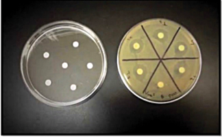
Colony forming units/ml
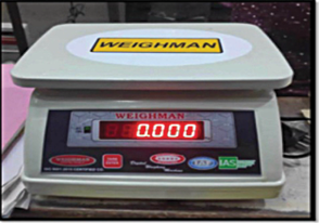
Electronic weighing machine Sensitivity: 1/1000 gm
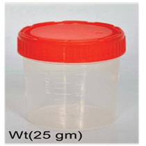
Tissue collecting container
Figure 2. Bacterial load sampling
NPWT dressings comprised of application of a sterile porous sponge pad with an open pore structure (400–600 microns) with transparent barrier dressing connected to the V.A.C.ULTA™ [2] Therapy System (3M-KCI USA, Inc) through a connecting channel. The exudates were collected in the canister attached to the VAC Machine. The negative pressure was maintained at 125 mm Hg in continuous mode. This entire dressing was kept for 7 days. The conventional dressings included daily dressing with washing with a copious amount of saline followed by Povidone Iodine 5% (Betadine® Win Medicare Limited) wash and Chlorhexidine impregnated Gauze dressings (Bactigras® Smith & Nephew Pvt Ltd) covered with rolls of sterile cotton pads gamzee soaked in 5% betadine tied over with sterile cotton bandage (Figure 3).

VAC DRESSING

CONVENTIONAL DRESSING
Figure 3. Types of dressings
On day seven the same measurements were repeated, and tissue was sent for bacterial load estimation. Pro-forma was given to patients to record date of admission, mode of injury, type of dressing applied, problems encountered and level of satisfaction (Figure 4). Patient compliance was ensured as all patients were admitted and were explained the procedures adequately.
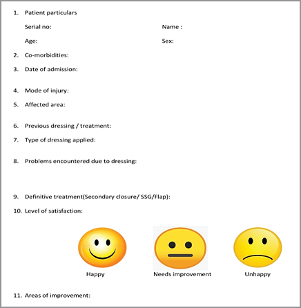
Figure 4. Patient proforma
The individual steps of the data extraction and risk-of-bias assessment procedures were always conducted by one person and checked by another; disagreements were resolved by consensus. All data were entered in a Microsoft Excel for Mac (version 16.63.1) spreadsheet which was prepared and validated for the data form. The statistical software SPSS 26 (IBM Corp. Released 2020. IBM SPSS Statistics for Windows, version 27.0. Armonk, NY: IBM Corp) was used for the analysis. Continuous variables were expressed as Mean, Median and Standard Deviation and compared across the groups using the Mann–Whitney U test. Categorical variables are expressed as the number of patients and percentage of patients and compared across the groups using Pearson’s Chi Square test for Independence of Attributes/Fisher’s Exact Test as appropriate. An alpha level of 5% has been taken and a p < 0.05 was considered significant.
Results
A total of 48 patients were enrolled as per the protocol (Figure 5) and participated in the study with a mean age of 46.83 (SD = 16.4) years (range 12–78 years). Most patients were in the age group 40–60 years of age (n = 21; 43.75%). The mean age in gNPWT was 46.38 (SD = 16.3) years and in gCD = 47.29 (SD = 16.35) years. There was a male preponderance of 3.8:1. The most common aetiology of the wound were Necrotising soft tissue infections 29.16% (n = 14) and soft tissue loss post lower limb compound fracture 29.16% (n = 14). Other aetiologies were post cellulitis raw area, post-burn ulcer and decubitus ulcer. Time of presentation since initial symptoms forms an essential component of wound healing. The mean time of presentation in both the groups (gNPWT = 15.58 (SD = 2.14) days; (gCD = 17.50 (SD = 2.17) days) showed no statistically significant difference. Before the initial debridement, the wound swab culture showed Klebsiella pneumoniae as the most common micro-organism 25% (n = 12) followed by 12.5% Pseudomonas aeruginosa (n = 6) and Staphylococcal species (n = 6). Rest micro-organisms included Gram-positive coccus, Gram-negative bacillus, anaerobes and facultative anaerobes. Most wounds showed a mixed pattern of involvement.
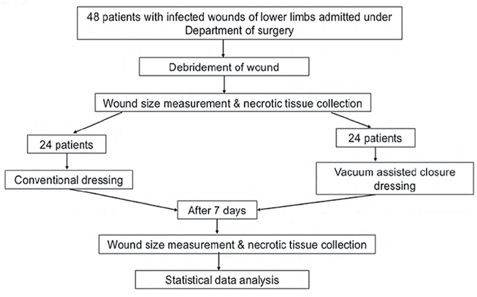
Figure 5. Study protocol
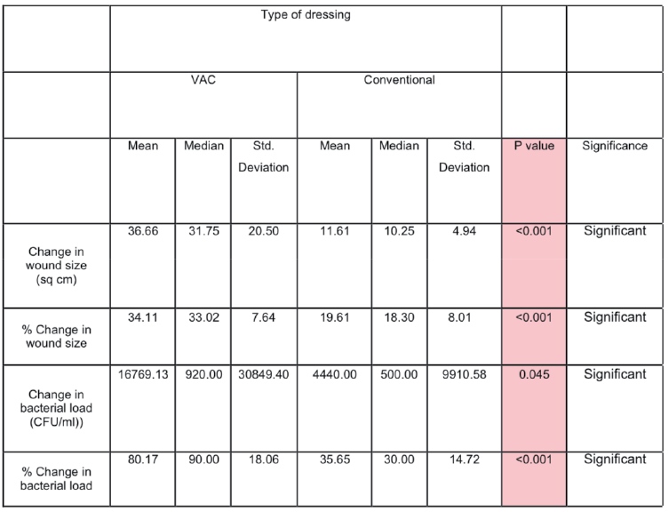
The mean wound area in gNPWT was 114.32 (SD = 68.3) cm2 and in gCD was 66.47 (SD = 30.71) cm2. No statistically significant difference was observed in the wound sizes in the two groups. The mean reduction in wound-area (∆MeanWA) was 36.66 (SD = 20.5) cm2 in the gNPWT and 11.61 (SD = 4.94) cm2 in the gCD (p < 0.05). Mean Percentage reduction in wound-area (∆Mean%WA) was 34.11% in the GNPWT and 19.61% in the gCD (p < 0.05). The mean bacterial load in gNPWT was 20773 (SD = 374.05) CFU/ml and in gCD was 12521 (SD = 279.61) CFU/ml. No statistically significant difference was observed in the wound bacterial load in the two dressing groups. The Mean difference in Wound-Bacterial-Load (∆MeanWBL) was 16769.13 CFU/ml in the gNPWT group and 4440 CFU/ml in the gCD group (p < 0.05). The Mean Percentage change in wound-bacterial-load (∆Mean%WBL) was 80.17% in the gNPWT and 35.65% in the gCD (p < 0.05) (Table 1).
Table 1. Primary outcomes
In the subset analysis of data ∆Mean%WA and ∆Mean%WBL were analysed for gender, Gram Stain, Anaerobic, aerobic, facultative anaerobic bacteria and type of wound. For ∆Mean%WA all parameters were statistically significant except female gender (p = 0.08), anaerobe bacteria (p = 0.22), Decubitus ulcers (p = 0.16), Cellulitis (p = 0.22) and post burn injury (p = 0. 22). Similarly ∆Mean%WBL all values were significant except anaerobe bacteria (p = 0.10), Decubitus ulcers (p = 0.06), Cellulitis (p = 0.22) and post burn injury (p = 0.22) (Table 2).
After the initial debridement in all patients, wound swab was taken using swab sticks, to assess the microbiological profile. The culture reports revealed 9 main varieties of bacteria amongst others. It was found that Klebsiella pneumoniae was isolated maximum number of times from the wounds (12 out of 48 patients) followed by Pseudomonas aeruginosa in 6 out of 48 patients and Staphylococcal species in 6 out of 48 patients. Rest of the microbiological profile is as shown below (Figure 6).
Table 2. Subset analysis
|
Sex |
% Change in |
% Change in |
||
|
MALE |
VAC |
Mean |
33.27 |
83.20 |
|
Median |
33.00 |
90.00 |
||
|
Std. deviation |
6.88 |
15.16 |
||
|
Conventional |
Mean |
16.77 |
38.75 |
|
|
Median |
14.85 |
40.00 |
||
|
Std. deviation |
5.36 |
15.44 |
||
|
|
p value |
<0.001 |
<0.001 |
|
|
Significance |
Significant |
Significant |
||
|
FEMALE |
VAC |
Mean |
40.01 |
60.00 |
|
Median |
36.00 |
50.00 |
||
|
Std. deviation |
11.76 |
26.46 |
||
|
Conventional |
Mean |
26.51 |
28.57 |
|
|
Median |
27.78 |
30.00 |
||
|
Std. deviation |
9.53 |
10.69 |
||
|
|
p value |
0.087 |
0.035 |
|
|
Significance |
Not significant |
Significant |
||
|
Organism isolated |
% Change in |
% Change in |
||
|
Gram positive coccus |
VAC |
Mean |
39.62 |
84.00 |
|
Median |
39.92 |
80.00 |
||
|
Std. deviation |
5.98 |
6.93 |
||
|
Conventional |
Mean |
13.04 |
30.00 |
|
|
Median |
12.16 |
30.00 |
||
|
Std. deviation |
2.69 |
10.00 |
||
|
|
p value |
0.025 |
0.023 |
|
|
Significance |
Significant |
Significant |
||
|
Gram negative bacillus |
VAC |
Mean |
33.57 |
79.60 |
|
Median |
32.41 |
90.00 |
||
|
Std. deviation |
7.75 |
19.23 |
||
|
Conventional |
Mean |
21.17 |
37.22 |
|
|
Median |
19.12 |
30.00 |
||
|
Std. deviation |
8.27 |
15.65 |
||
|
|
p value |
<0.001 |
<0.001 |
|
|
Significance |
Significant |
Significant |
||
|
Aerobic/anaerobic organism |
% Change in |
% Change in |
||
|
Aerobe |
VAC |
Mean |
33.50 |
79.73 |
|
Median |
33.04 |
80.00 |
||
|
Std. deviation |
6.87 |
16.94 |
||
|
Conventional |
Mean |
18.98 |
30.00 |
|
|
Median |
17.58 |
30.00 |
||
|
Std. deviation |
8.75 |
11.18 |
||
|
|
p value |
0.003 |
<0.001 |
|
|
Significance |
Significant |
Significant |
||
|
Facultative |
VAC |
Mean |
32.97 |
83.50 |
|
Median |
32.36 |
91.00 |
||
|
Std. deviation |
6.92 |
16.46 |
||
|
Conventional |
Mean |
20.53 |
37.50 |
|
|
Median |
18.82 |
30.00 |
||
|
Std. deviation |
8.38 |
16.58 |
||
|
|
p value |
0.003 |
<0.001 |
|
|
Significance |
Significant |
Significant |
||
|
Aerobe & anaerobe |
VAC |
Mean |
46.07 |
66.00 |
|
Median |
46.07 |
66.00 |
||
|
Std. deviation |
10.15 |
36.77 |
||
|
Conventional |
Mean |
14.50 |
50.00 |
|
|
Median |
14.50 |
50.00 |
||
|
Std. deviation |
0.50 |
0.00 |
||
|
|
p value |
<0.001 |
0.102 |
|
|
Significance |
Significant |
Not significant |
||
|
Type of wound |
% Change in |
% Change in |
||
|---|---|---|---|---|
|
COMPOUND FRACTURE |
VAC |
Mean |
31.37 |
77.50 |
|
Median |
33.00 |
80.00 |
||
|
Std. deviation |
6.11 |
20.62 |
||
|
Conventional |
Mean |
19.43 |
35.00 |
|
|
Median |
19.12 |
35.00 |
||
|
Std. deviation |
5.54 |
12.91 |
||
|
|
p value |
0.028 |
0.028 |
|
|
Significance |
Significant |
Significant |
||
|
NSTI |
VAC |
Mean |
33.62 |
82.43 |
|
Median |
31.82 |
90.00 |
||
|
Std. deviation |
7.99 |
19.27 |
||
|
Conventional |
Mean |
15.80 |
38.57 |
|
|
Median |
14.85 |
40.00 |
||
|
Std. deviation |
3.20 |
9.00 |
||
|
|
p value |
0.002 |
0.002 |
|
|
Significance |
Significant |
Significant |
||
|
Median |
25.00 |
50.00 |
||
|
Std. deviation |
|
|
||
|
|
p value |
NA |
NA |
|
|
Significance |
NA |
NA |
||
|
DEVITALIZING INJURIES |
VAC |
Mean |
37.50 |
83.75 |
|
Median |
38.29 |
80.00 |
||
|
Std. deviation |
2.78 |
7.50 |
||
|
Conventional |
Mean |
20.88 |
36.00 |
|
|
Median |
26.15 |
40.00 |
||
|
Std. deviation |
9.99 |
15.17 |
||
|
|
p value |
0.014 |
0.012 |
|
|
Significance |
Significant |
Significant |
||
|
DECUBITUS ULCER |
VAC |
Mean |
44.63 |
65.00 |
|
Median |
44.63 |
65.00 |
||
|
Std. deviation |
12.20 |
35.36 |
||
|
Conventional |
Mean |
28.21 |
27.50 |
|
|
Median |
27.16 |
30.00 |
||
|
Std. deviation |
11.02 |
5.00 |
||
|
|
p value |
0.165 |
0.059 |
|
|
Significance |
Not Significant |
Not Significant |
||
|
CELLULITIS |
VAC |
Mean |
26.67 |
94.00 |
|
Median |
26.67 |
94.00 |
||
|
Std. deviation |
|
|
||
|
Conventional |
Mean |
16.13 |
50.00 |
|
|
Median |
16.13 |
50.00 |
||
|
Std. deviation |
7.65 |
42.43 |
||
|
|
p value |
0.221 |
0.221 |
|
|
Significance |
Not Significant |
Not Significant |
||
|
POST BURNS |
VAC |
Mean |
31.65 |
93.00 |
|
Median |
31.65 |
93.00 |
||
|
Std. deviation |
0.11 |
1.41 |
||
|
Conventional |
Mean |
13.46 |
20.00 |
|
|
Median |
13.46 |
20.00 |
||
|
Std. deviation |
NA |
NA |
||
|
|
p value |
0.221 |
0.221 |
|
|
Significance |
Not Significant |
Not Significant |
||
|
OTHERS |
VAC |
Mean |
36.13 |
81.00 |
|
Median |
36.13 |
81.00 |
||
|
Std. deviation |
13.19 |
15.56 |
||
|
Conventional |
Mean |
38.00 |
20% |
|
|
Median |
38.00 |
20% |
||
|
Std. deviation |
NA |
NA |
||
|
|
p value |
1.03 |
0.024 |
|
|
Significance |
Not Significant |
Significant |
||

Figure 6. Multi-resistant microorganisms isolated
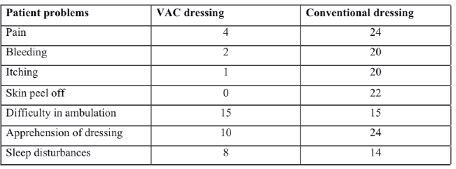
The secondary outcome was evaluated in terms of problems encountered by patients in either of the two dressings. Patients complained of local pain, bleeding, itching, skin discolouration, apprehension of dressing and sleep disturbances during the therapy period. The problems encountered were found to be less in gNPWT. The results are tabulated in Table 3.
Table 3. Secondary outcomes
Discussion
Infected wounds of the lower limbs form a significant portion of the problem statement, contributing to the morbidity amongst patients in very high numbers. The actual burden is formed by chronic infected lower limb wounds that do not have any arteriovenous compromise, but have no standardised methodology of management, leading to ambiguous and under confident treatment. This would not only compromise on the patient recovery but also involve unmonitored yet substantial utilisation of hospital infrastructure and resources.
In this study, we plan to address this problem statement, by observing the clinico-microbiological differences between infected lower limb wounds treated with NPWT and conventional dressings. At the end of the study, we observed that among 48 patients with infected chronic lower limb wounds treated with NPWT had better healing (change in wound size and bacterial load) as compared to conventional dressings. The patients were first analysed for any differences between the two dressing groups. There were no significant differences found in the distribution of patients in the two dressing groups as per age, gender, co-morbidities, time of presentation and wound aetiology. A statistically significant difference was observed in the mean wound size reduction and mean decrease in bacterial load in NPWT dressing as compared to conventional dressings, thus showing that VAC dressings are more effective than conventional dressings.
Joseph et al. [2] in 2000, conducted a prospective randomised controlled trial with 24 patients and 36 wounds (chronic wounds present for more than 1 month: post amputation stumps; infected wounds) 111 amongst which half received NPWT and other half received saline dressings. Significant difference in percent change in wound volume was observed at 6 weeks in favour of NPWT (78 vs. 30%). It was a blinded study which concluded that NPWT has a positive impact on wound healing. Wongworawat et al. [3] in 2003 conducted a prospective study with 14 patients having orthopaedic infected wounds all >20 cm2 on lower limbs. The observed average wound size reduction was 43% in 10 days. They therefore concluded that NPWT enhances wound reduction and is advantageous in managing infected wounds.
In the Multicentric UK WOLLF RCT 2018 (UK Wound management of Lower Limb Fractures) for assessing the merit of NPWT with standard dressing for soft tissue raw areas post lower limb fracture the study concluded that there is no added advantage of NPWT. The study was conducted among 460 patients (NPWT; n = 226 and standard dressings n = 234) and showed no difference in surgical site infections or other complications at any point in the 12 month postop period. Also NPWT was associated with increased cost of treatment [4]. Costa et al. [5] in the recent 2020 WHiST RCT conducted among 1548 patients with major trauma to lower limb concluded that there is no added advantage of NPWT which contradicts our findings. Their main limitation was the use of NPWT in an emergency setting and many patients were lost to follow-up. But Benrashid [6] 2020 study concluded that NPWT usage reduced the lower limb infection rate. In 504 patients randomised into two groups (NPWT; n = 225 and standard dressings n = 279) NPWT reduced infection with odds ratio 0.32; confidence interval 0.17–0.63; and p < 0.01.
Yang et al. [7] demonstrated in their 53 patients with chronic venous ulcer wounds, the wound healing rate on day 7 for NPWT (33±10%) was statistically significant than regular dressing group (24±9%) with p < 0.05 and increased to even better on day 14 with p < 0.001. A similar improvement was also seen in the transcutaneous oxygen saturation post NPWT in toes compared to normal dressing with p < 0.05 on day 7 and p < 0.001 on day 14. This study also concluded that cyclic application of NPWT has better outcome compared to single pressure setting in setting of lower limb chronic venous ulcers.
Similar results were shown in the series by Yao et al. [8] in chronic lower extremity ulcers. In this cohort of 342 patients managed equally with NPWT and Conventional dressings and found that NPWT patients were 2.63 times (95% CI = 1.87–3.70) more likely to have wound closure. This study showed that irrespective of comorbidity status NPWT had better wound healing than normal dressings. Moues et al. in 2004 studied 54 patients who needed open wound management before surgical closure. They were randomised in to two groups (NPWT; n = 29 and conventional moist gauze therapy n = 25). Wound surface area reduction was significantly larger in vacuum-assisted closure-treated wounds: 3.8±0.5 %/day compared to conventional-treated wounds (1.7±0.6 %/day; p < 0.05). The total quantitative bacterial load was equal in both groups. However, nonfermentive gram negative bacilli showed a significant decrease in vacuum-assisted closure-treated wounds (p < 0.05), whereas Staphylococcus aureus showed a significant increase in vacuum-assisted closure-treated wounds (p < 0.05). However, studies also states higher bacterial load in sponge used for NPWT. Yusuf et al. [9] in their series of 68 samples found that the samples had a high load as 1,000 to 1,00,000 CFU/gm of foam. Moong et al. [10] demonstrated results of 704 microbiological swabs from 97 patients in 110 wound localizations in a mono-centric, retrospective study and concluded that NPWT did not improve bacterial bioburden in 77% of cases and the duration of NPWT did not affect the result. Furthermore, no significant effect of NPWT could be found for either anaerobic (p = 0.96) or aerobic bacteria (p = 0.43).
In a systematic review by Glass et al. [11] in 2017, included 8 studies using conventional NPWT, that failed to demonstrate an observable effect on bacterial load, 7 studies reported that NPWT was inherently bacteriostatic and 5 others reported species selectivity with suppression of nonfermentive gram-negative bacilli (NFGNB), including Pseudomonas sp. Enescu et al. [12] in 2020 demonstrated the effective ness of NPWT dressings in children. This study retrospectively analysed nine children with severe ankle and foot injury with mean age of 10 years (range 3 years 5 months to 14 years 4 months). The study evaluated data from 2016 to 2019. NPWT has resulted in excellent results both in short and long term. This was safe, easy to use with less dressing changes, decreased pain and anxiety and resulted in better well granulated contracted wound facilitating split skin graft in all but one patient. Similar results were shown by Masumota et al. [13] in 6 children with lower limb surgical site infection. A comparative study by Kumar et al. [14] in 2021, concluded that the average reduction in wound size from day 1 to day 21 was statistically significant, with the NPWT group showing more rapid reduction in wound size compared to conventional dressing group (35% in NPWT) as compared to 17% in Group B (conventional dressing) (p < 0.001).
Strengths of this study were as follows: 1) it was conducted at a tertiary care centre with well-equipped infrastructure; 2) all the patients were admitted in the hospital, therefore ensuring good compliance and this also helped in adequate monitoring of patients, should any complications arise; 3) this study encompasses a plethora of variables, that affect the process of wound healing and has done adequate justice by comparing them under one roof, that has not been accomplished in previous studies; 4) all the patients were distributed amongst the two groups in a manner that, bias related to demographic profile of patients, co-morbidities and wound characters were avoided; 5) there was no discrepancy in terms of wound sizes and infectivity, in the two groups, that further enhances the quality of this study; 6) the results have shown statistically significant data, which was tabulated using appropriate analytical tools; through this study, we have been able to infer association between the continuous variables (time of presentation, isolated organisms and type of wound) and type of dressing used in terms of the primary outcomes; 7) the study has also given the opportunity to derive the secondary outcome (patient problems) which have further broadened the horizon of NPWT and this aspect can be studied better in successive studies.
The limitations of this study were: 1) single centre study; 2) relatively smaller sample size that might affect the generalisability of the study; 3) the measurement of wound sizes may be objectified by using technically advanced tools to yield better results; 4) no growth was seen in two patients on pus culture, which may have had an impact on the results; 5) in patients presenting within 15–21 days, primary outcomes could not be compared as there were no patients in the conventional dressing group, presenting within this time frame.
Conclusion
The effectiveness of NPWT in promoting wound healing has been widely accepted by surgeons but the number of advanced studies demonstrating its effectiveness is limited for standardised management of infected lower limb wounds. This study was conducted in view of scarcity of high-quality data. Evidence indicated that the effectiveness of such therapy is at least as good as or better than current local treatment for wounds. Through this study, we have observed that the NPWT has been instrumental in statistically significant reduction of wound size as well as bacterial load in infected wounds of lower limbs. It also brings out the variable effects of NPWT in a particular type of wound or organism isolated as evident from the subgroup analysis. These parameters can be explored more in further studies in future.
Provenance and peer review. Not commissioned, externally peer-reviewed.
Conflict of interest declaration. The authors declare that they have no affiliations with or involvement in any organisation or entity with any financial interest in the subject matter or materials used in this manuscript.
Funding statement. N/A. No funding institutions were involved.
Consent. A written and informed consent was obtained from all the subjects in the below mentioned format.
Author contributions
Onkar Singh – concept and design, critical revision, final approval of the version to be published.
Aprajita Atreya – literature collection and data search, drafting of manuscript, data analysis and interpretation.
Vipin Venugopal Nair – supervised the research and critically revised the manuscript.
Anannya Dutta – provided overall supervision, final approval of the version to be published.
Sunil Kumar – literature collection and data search.
References
1. Petkar KS, Dhanraj P, Kingsly PM, Sreekar H, Lakshmanarao A, Lamba S, Shetty R, Zachariah JR. A prospective randomized controlled trial comparing negative pressure dressing and conventional dressing methods on split-thickness skin grafts in burned patients. Burns 2011; 37(6): 925–929.
2. Joseph E, Hamori CA, Bergman S, Roaf E, Swann NF, Anastasi GW. A prospective randomised trial of vacuum assisted closure versus standard therapy of chronic nonhealing wounds. Wounds 2000; 12: 60–67.
3. Wongworawat MD, Schnall SB, Holtom PD, Moon C, Schiller F. Negative pressure dressings as an alternative technique for the treatment of infected wounds. Clin Orthop Relat Res 2003; 414: 45–48.
4. Costa ML, Achten J, Bruce J, Davis S, Hennings S, Willett K, Petrou S, Jeffery S, Griffin D, Parker B, Masters J, Lamb SE, Tutton E, Parsons N. Negative-pressure wound therapy versus standard dressings for adults with an open lower limb fracture: The WOLLF RCT. Health Technol Assess 2018; 22(73): 1–162.
5. Costa ML, Achten J, Knight R, Png ME, Bruce J, Dutton S, Madan J, Vadher K, Dritsaki M, Masters J, Spoors L, Campolier M, Parsons N, Fernandez M, Jones S, Grant R, Nanchahal. Negative-pressure wound therapy compared with standard dressings following surgical treatment of major trauma to the lower limb: The WHiST RCT. Health Technol Assess 2020; 24(38): 1–86.
6. Benrashid E, Youngwirth LM, Guest K, Cox MW, Shortell CK, Dillavou ED. Negative pressure wound therapy reduces surgical site infections. J Vasc Surg 2020; 71(3): 896–904.
7. Yang ML, Zhou XJ, Zhu YG, Jiang DL, Ding LT, Chu GP, Zhao P, Cheng J, Lyu GZ, Li QF. Clinical efficacy and influencing factors of different modes of continuous negative pressure wound therapy on venous ulcer wounds of lower limbs. Zhonghua Shao Shang Za Zhi 2020; 36(12): 1149–1158.
8. Yao M, Fabbi M, Hayashi H, Park N, Attala K, Gu G, French MA, Driver VR. A retrospective cohort study evaluating efficacy in high-risk patients with chronic lower extremity ulcers treated with negative pressure wound therapy. Int Wound J 2014; 11(5): 483–488.
9. Yusuf E, Jordan X, Clauss M, Borens O, Mäder M, Trampuz A. High bacterial load in negative pressure wound therapy (NPWT) foams used in the treatment of chronic wounds. Wound Repair Regen 2013; 21(5): 677–681.
10. Moog P, Jensch M, Betzl J, Bauer AT, Cerny MK, Schmauss D, Kükrek H, Erne H, Machens HG, Megerle K. Bacterial bioburden of wounds: Influence of debridement and negative-pressure wound therapy (NPWT). J Wound Care 2021; 30(8): 604–611.
11. Glass GE, Murphy GRF, Nanchahal J. Does negative pressure wound therapy influence subjacent bacterial growth? A systematic review. J Plast Reconstr Aesthet Surg 2017; 70(8): 1028–1037.
12. Enescu DM, Stoicescu S, Tomiţă M, Nacea I, Ioniţă D, Tatar R. Management of lower extremity traumatic injuries with negative pressure wound therapy: Experience of a Pediatric Plastic Surgery Department. Injury 2020; 51: S9–S15.
13. Masumoto K, Nagata K, Oka Y, Kai H, Yamaguchi S, Wada M, Kusuda T, Hara T, Hirose S, Iwasaki A, Taguchi T. Successful treatment of an infected wound in infants by a combination of negative pressure wound therapy and arginine supplementation. Nutrition 2011; 27(11–12): 1141–1145.
14. Kumar M, Kuamr A, Ahmad ME. Comparative study of negative pressure wound therapy versus conventional wound therapy of lower limb ulcers. Int J Heal Clin Res 2021; 4(1): 252–256.